5J5Q

AMP-PNP-stabilized ATPase domain of topoisomerase IV from Streptococcus pneumoniae, complex type II


Experimental Data Snapshot

  • Method: X-RAY DIFFRACTION
  • Resolution: 2.83 Å
  • R-Value Free: 0.263 
  • R-Value Work: 0.184 
  • R-Value Observed: 0.188 

wwPDB Validation   3D Report Full Report


Ligand Structure Quality Assessment 


This is version 1.2 of the entry. See complete history


Literature

Trapping of the transport-segment DNA by the ATPase domains of a type II topoisomerase.

Laponogov, I.Pan, X.S.Veselkov, D.A.Skamrova, G.B.Umrekar, T.R.Fisher, L.M.Sanderson, M.R.

(2018) Nat Commun 9: 2579-2579

  • DOI: https://doi.org/10.1038/s41467-018-05005-x
  • Primary Citation of Related Structures:  
    5J5P, 5J5Q

  • PubMed Abstract: 

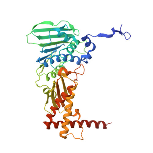
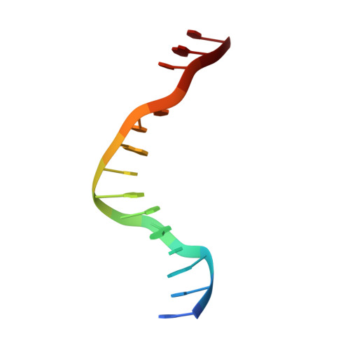
    Type II topoisomerases alter DNA topology to control DNA supercoiling and chromosome segregation and are targets of clinically important anti-infective and anticancer therapeutics. They act as ATP-operated clamps to trap a DNA helix and transport it through a transient break in a second DNA. Here, we present the first X-ray crystal structure solved at 2.83 Å of a closed clamp complete with trapped T-segment DNA obtained by co-crystallizing the ATPase domain of S. pneumoniae topoisomerase IV with a nonhydrolyzable ATP analogue and 14-mer duplex DNA. The ATPase dimer forms a 22 Å protein hole occupied by the kinked DNA bound asymmetrically through positively charged residues lining the hole, and whose mutagenesis impacts the DNA decatenation, DNA relaxation and DNA-dependent ATPase activities of topo IV. These results and a side-bound DNA-ParE structure help explain how the T-segment DNA is captured and transported by a type II topoisomerase, and reveal a new enzyme-DNA interface for drug discovery.


  • Organizational Affiliation

    Randall Centre for Cell and Molecular Biophysics, 3rd Floor New Hunt's House, Faculty of Life Sciences and Medicine, King's College London, London, SE1 1UL, UK.


Macromolecules

Find similar proteins by:  (by identity cutoff)  |  3D Structure
Entity ID: 1
MoleculeChains Sequence LengthOrganismDetailsImage
DNA topoisomerase 4 subunit B
A, B, C, D
409Streptococcus pneumoniaeMutation(s): 0 
Gene Names: parESP_0852
EC: 5.99.1.3
UniProt
Find proteins for Q59961 (Streptococcus pneumoniae serotype 4 (strain ATCC BAA-334 / TIGR4))
Explore Q59961 
Go to UniProtKB:  Q59961
Entity Groups  
Sequence Clusters30% Identity50% Identity70% Identity90% Identity95% Identity100% Identity
UniProt GroupQ59961
Sequence Annotations
Expand
  • Reference Sequence

Find similar nucleic acids by:  Sequence   |   3D Structure  

Entity ID: 2
MoleculeChains LengthOrganismImage
DNA (5'-D(*GP*CP*AP*TP*AP*TP*AP*TP*AP*TP*AP*TP*GP*C)-3')
E, F
14synthetic construct
Sequence Annotations
Expand
  • Reference Sequence
Experimental Data & Validation

Experimental Data

  • Method: X-RAY DIFFRACTION
  • Resolution: 2.83 Å
  • R-Value Free: 0.263 
  • R-Value Work: 0.184 
  • R-Value Observed: 0.188 
  • Space Group: C 1 2 1
Unit Cell:
Length ( Å )Angle ( ˚ )
a = 124.95α = 90
b = 72.74β = 91.89
c = 222.96γ = 90
Software Package:
Software NamePurpose
PHENIXrefinement
xia2data reduction
xia2data scaling
PHASERphasing

Structure Validation

View Full Validation Report


Ligand Structure Quality Assessment 


Entry History & Funding Information

Deposition Data


Funding OrganizationLocationGrant Number
Biotechnology and Biological Sciences Research CouncilUnited KingdomBB/K010069/1

Revision History  (Full details and data files)

  • Version 1.0: 2017-07-26
    Type: Initial release
  • Version 1.1: 2018-07-18
    Changes: Data collection, Database references
  • Version 1.2: 2024-01-10
    Changes: Data collection, Database references, Derived calculations, Refinement description