5IWQ

Crystal structure of aspartate aminotransferase (AspAT) from Corynebacterium glutamicum ATCC 13032


Experimental Data Snapshot

  • Method: X-RAY DIFFRACTION
  • Resolution: 2.00 Å
  • R-Value Free: 0.199 
  • R-Value Work: 0.148 
  • R-Value Observed: 0.151 

wwPDB Validation   3D Report Full Report


Ligand Structure Quality Assessment 


This is version 1.1 of the entry. See complete history


Literature

Structural Insights into a Novel Class of Aspartate Aminotransferase from Corynebacterium glutamicum.

Son, H.F.Kim, K.J.

(2016) PLoS One 11: e0158402-e0158402

  • DOI: https://doi.org/10.1371/journal.pone.0158402
  • Primary Citation of Related Structures:  
    5IWQ

  • PubMed Abstract: 

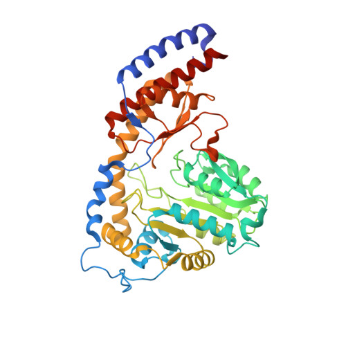
    Aspartate aminotransferase from Corynebacterium glutamicum (CgAspAT) is a PLP-dependent enzyme that catalyzes the production of L-aspartate and α-ketoglutarate from L-glutamate and oxaloacetate in L-lysine biosynthesis. In order to understand the molecular mechanism of CgAspAT and compare it with those of other aspartate aminotransferases (AspATs) from the aminotransferase class I, we determined the crystal structure of CgAspAT. CgAspAT functions as a dimer, and the CgAspAT monomer consists of two domains, the core domain and the auxiliary domain. The PLP cofactor is found to be bound to CgAspAT and stabilized through unique residues. In our current structure, a citrate molecule is bound at the active site of one molecule and mimics binding of the glutamate substrate. The residues involved in binding of the PLP cofactor and the glutamate substrate were confirmed by site-directed mutagenesis. Interestingly, compared with other AspATs from aminotransferase subgroup Ia and Ib, CgAspAT exhibited unique binding sites for both cofactor and substrate; moreover, it was found to have unusual structural features in the auxiliary domain. Based on these structural differences, we propose that CgAspAT does not belong to either subgroup Ia or Ib, and can be categorized into a subgroup Ic. The phylogenetic tree and RMSD analysis also indicates that CgAspAT is located in an independent AspAT subgroup.


  • Organizational Affiliation

    School of Life Sciences, KNU Creative BioResearch Group, Kyungpook National University, Daehak-ro 80, Buk-ku, Daegu 702-701, Korea.


Macromolecules
Find similar proteins by:  (by identity cutoff)  |  3D Structure
Entity ID: 1
MoleculeChains Sequence LengthOrganismDetailsImage
ASPARTATE AMINOTRANSFERASE
A, B
434Corynebacterium glutamicum ATCC 13032Mutation(s): 0 
Gene Names: aspBcg0294
EC: 2.6.1.1
UniProt
Find proteins for Q8NTR2 (Corynebacterium glutamicum (strain ATCC 13032 / DSM 20300 / BCRC 11384 / JCM 1318 / LMG 3730 / NCIMB 10025))
Explore Q8NTR2 
Go to UniProtKB:  Q8NTR2
Entity Groups  
Sequence Clusters30% Identity50% Identity70% Identity90% Identity95% Identity100% Identity
UniProt GroupQ8NTR2
Sequence Annotations
Expand
  • Reference Sequence
Small Molecules
Ligands 3 Unique
IDChains Name / Formula / InChI Key2D Diagram3D Interactions
PLP
Query on PLP

Download Ideal Coordinates CCD File 
C [auth A],
I [auth B]
PYRIDOXAL-5'-PHOSPHATE
C8 H10 N O6 P
NGVDGCNFYWLIFO-UHFFFAOYSA-N
FLC
Query on FLC

Download Ideal Coordinates CCD File 
J [auth B]CITRATE ANION
C6 H5 O7
KRKNYBCHXYNGOX-UHFFFAOYSA-K
GOL
Query on GOL

Download Ideal Coordinates CCD File 
D [auth A]
E [auth A]
F [auth A]
G [auth A]
H [auth A]
D [auth A],
E [auth A],
F [auth A],
G [auth A],
H [auth A],
K [auth B],
L [auth B],
M [auth B],
N [auth B],
O [auth B]
GLYCEROL
C3 H8 O3
PEDCQBHIVMGVHV-UHFFFAOYSA-N
Experimental Data & Validation

Experimental Data

  • Method: X-RAY DIFFRACTION
  • Resolution: 2.00 Å
  • R-Value Free: 0.199 
  • R-Value Work: 0.148 
  • R-Value Observed: 0.151 
  • Space Group: C 1 2 1
Unit Cell:
Length ( Å )Angle ( ˚ )
a = 103.388α = 90
b = 53.521β = 104.23
c = 170.224γ = 90
Software Package:
Software NamePurpose
REFMACrefinement
HKL-2000data collection
PDB_EXTRACTdata extraction
HKL-2000data reduction
HKL-2000data scaling
HKL-2000phasing

Structure Validation

View Full Validation Report


Ligand Structure Quality Assessment 


Entry History 

Deposition Data

Revision History  (Full details and data files)

  • Version 1.0: 2016-07-20
    Type: Initial release
  • Version 1.1: 2023-11-08
    Changes: Data collection, Database references, Derived calculations, Refinement description