5HPJ

Photobacterium profundum alpha-carbonic anhydrase


Experimental Data Snapshot

  • Method: X-RAY DIFFRACTION
  • Resolution: 1.50 Å
  • R-Value Free: 0.168 
  • R-Value Work: 0.139 
  • R-Value Observed: 0.141 

wwPDB Validation   3D Report Full Report


This is version 1.4 of the entry. See complete history


Literature

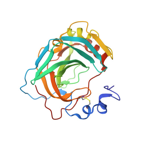
A High-Resolution Crystal Structure of a Psychrohalophilic alpha-Carbonic Anhydrase from Photobacterium profundum Reveals a Unique Dimer Interface.

Somalinga, V.Buhrman, G.Arun, A.Rose, R.B.Grunden, A.M.

(2016) PLoS One 11: e0168022-e0168022

  • DOI: https://doi.org/10.1371/journal.pone.0168022
  • Primary Citation of Related Structures:  
    5HPJ

  • PubMed Abstract: 

    Bacterial α-carbonic anhydrases (α-CA) are zinc containing metalloenzymes that catalyze the rapid interconversion of CO2 to bicarbonate and a proton. We report the first crystal structure of a pyschrohalophilic α-CA from a deep-sea bacterium, Photobacterium profundum. Size exclusion chromatography of the purified P. profundum α-CA (PprCA) reveals that the protein is a heterogeneous mix of monomers and dimers. Furthermore, an "in-gel" carbonic anhydrase activity assay, also known as protonography, revealed two distinct bands corresponding to monomeric and dimeric forms of PprCA that are catalytically active. The crystal structure of PprCA was determined in its native form and reveals a highly conserved "knot-topology" that is characteristic of α-CA's. Similar to other bacterial α-CA's, PprCA also crystallized as a dimer. Furthermore, dimer interface analysis revealed the presence of a chloride ion (Cl-) in the interface which is unique to PprCA and has not been observed in any other α-CA's characterized so far. Molecular dynamics simulation and chloride ion occupancy analysis shows 100% occupancy for the Cl- ion in the dimer interface. Zinc coordinating triple histidine residues, substrate binding hydrophobic patch residues, and the hydrophilic proton wire residues are highly conserved in PprCA and are identical to other well-studied α-CA's.


  • Organizational Affiliation

    Department of Plant and Microbial Biology, North Carolina State University, Raleigh, NC, United States of America.


Macromolecules
Find similar proteins by:  (by identity cutoff)  |  3D Structure
Entity ID: 1
MoleculeChains Sequence LengthOrganismDetailsImage
Carbonic anhydrase217Photobacterium profundum SS9Mutation(s): 0 
Gene Names: EBIG1812PBPRA3376
UniProt
Find proteins for Q6LM17 (Photobacterium profundum (strain SS9))
Explore Q6LM17 
Go to UniProtKB:  Q6LM17
Entity Groups  
Sequence Clusters30% Identity50% Identity70% Identity90% Identity95% Identity100% Identity
UniProt GroupQ6LM17
Sequence Annotations
Expand
  • Reference Sequence
Experimental Data & Validation

Experimental Data

  • Method: X-RAY DIFFRACTION
  • Resolution: 1.50 Å
  • R-Value Free: 0.168 
  • R-Value Work: 0.139 
  • R-Value Observed: 0.141 
  • Space Group: P 32 2 1
Unit Cell:
Length ( Å )Angle ( ˚ )
a = 60.461α = 90
b = 60.461β = 90
c = 95.464γ = 120
Software Package:
Software NamePurpose
PHENIXrefinement
DENZOdata reduction
SCALEPACKdata scaling
PHASERphasing
PDB_EXTRACTdata extraction
HKL-2000data reduction

Structure Validation

View Full Validation Report


Entry History & Funding Information

Deposition Data


Funding OrganizationLocationGrant Number
National Science Foundation (NSF, United States)United States1332341

Revision History  (Full details and data files)

  • Version 1.0: 2016-12-07
    Type: Initial release
  • Version 1.1: 2016-12-28
    Changes: Database references
  • Version 1.2: 2017-09-27
    Changes: Author supporting evidence
  • Version 1.3: 2019-11-27
    Changes: Author supporting evidence
  • Version 1.4: 2023-09-27
    Changes: Data collection, Database references, Refinement description