5F93

Blood group antigen binding adhesin BabA of Helicobacter pylori strain A730 in complex with blood group H Lewis b hexasaccharide


Experimental Data Snapshot

  • Method: X-RAY DIFFRACTION
  • Resolution: 2.99 Å
  • R-Value Free: 0.234 
  • R-Value Work: 0.181 
  • R-Value Observed: 0.184 

wwPDB Validation   3D Report Full Report


This is version 2.1 of the entry. See complete history


Literature

Structural Insights into Polymorphic ABO Glycan Binding by Helicobacter pylori.

Moonens, K.Gideonsson, P.Subedi, S.Bugaytsova, J.Romao, E.Mendez, M.Norden, J.Fallah, M.Rakhimova, L.Shevtsova, A.Lahmann, M.Castaldo, G.Brannstrom, K.Coppens, F.Lo, A.W.Ny, T.Solnick, J.V.Vandenbussche, G.Oscarson, S.Hammarstrom, L.Arnqvist, A.Berg, D.E.Muyldermans, S.Boren, T.Remaut, H.

(2016) Cell Host Microbe 19: 55-66

  • DOI: https://doi.org/10.1016/j.chom.2015.12.004
  • Primary Citation of Related Structures:  
    5F7K, 5F7L, 5F7M, 5F7N, 5F7W, 5F7Y, 5F8Q, 5F8R, 5F93, 5F97, 5F9A, 5F9D

  • PubMed Abstract: 

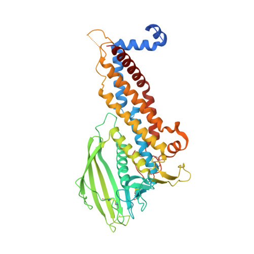
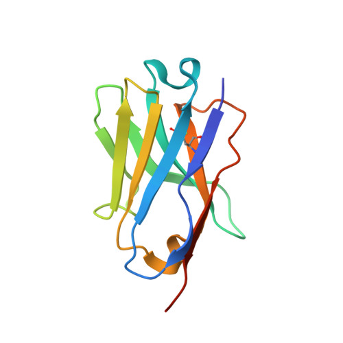
    The Helicobacter pylori adhesin BabA binds mucosal ABO/Le(b) blood group (bg) carbohydrates. BabA facilitates bacterial attachment to gastric surfaces, increasing strain virulence and forming a recognized risk factor for peptic ulcers and gastric cancer. High sequence variation causes BabA functional diversity, but the underlying structural-molecular determinants are unknown. We generated X-ray structures of representative BabA isoforms that reveal a polymorphic, three-pronged Le(b) binding site. Two diversity loops, DL1 and DL2, provide adaptive control to binding affinity, notably ABO versus O bg preference. H. pylori strains can switch bg preference with single DL1 amino acid substitutions, and can coexpress functionally divergent BabA isoforms. The anchor point for receptor binding is the embrace of an ABO fucose residue by a disulfide-clasped loop, which is inactivated by reduction. Treatment with the redox-active pharmaceutic N-acetylcysteine lowers gastric mucosal neutrophil infiltration in H. pylori-infected Le(b)-expressing mice, providing perspectives on possible H. pylori eradication therapies.


  • Organizational Affiliation

    Structural and Molecular Microbiology, Structural Biology Research Center, VIB, Pleinlaan 2, 1050 Brussels, Belgium; Structural Biology Brussels, Vrije Universiteit Brussel, Pleinlaan 2, 1050 Brussels, Belgium.


Macromolecules
Find similar proteins by:  (by identity cutoff)  |  3D Structure
Entity ID: 1
MoleculeChains Sequence LengthOrganismDetailsImage
Adhesin binding fucosylated histo-blood group antigen,Adhesin,Adhesin binding fucosylated histo-blood group antigenA,
B,
C [auth E],
D [auth G]
469Helicobacter pyloriMutation(s): 0 
Gene Names: babA2babA
UniProt
Find proteins for Q6DSX7 (Helicobacter pylori)
Explore Q6DSX7 
Go to UniProtKB:  Q6DSX7
Find proteins for O52269 (Helicobacter pylori)
Explore O52269 
Go to UniProtKB:  O52269
Entity Groups  
Sequence Clusters30% Identity50% Identity70% Identity90% Identity95% Identity100% Identity
UniProt GroupsO52269Q6DSX7
Sequence Annotations
Expand
  • Reference Sequence
Find similar proteins by:  (by identity cutoff)  |  3D Structure
Entity ID: 2
MoleculeChains Sequence LengthOrganismDetailsImage
Nanobody Nb-ER19E [auth C],
F [auth D],
G [auth F],
H
120Lama glamaMutation(s): 0 
Entity Groups  
Sequence Clusters30% Identity50% Identity70% Identity90% Identity95% Identity100% Identity
Sequence Annotations
Expand
  • Reference Sequence
Oligosaccharides

Help

Entity ID: 3
MoleculeChains Length2D Diagram Glycosylation3D Interactions
alpha-L-fucopyranose-(1-2)-beta-D-galactopyranose-(1-3)-[alpha-L-fucopyranose-(1-4)]2-acetamido-2-deoxy-beta-D-glucopyranose-(1-3)-beta-D-galactopyranose-(1-4)-alpha-D-glucopyranose
I, J, K, L
6N/A
Glycosylation Resources
GlyTouCan:  G30212HV
GlyCosmos:  G30212HV
GlyGen:  G30212HV
Experimental Data & Validation

Experimental Data

  • Method: X-RAY DIFFRACTION
  • Resolution: 2.99 Å
  • R-Value Free: 0.234 
  • R-Value Work: 0.181 
  • R-Value Observed: 0.184 
  • Space Group: P 1 21 1
Unit Cell:
Length ( Å )Angle ( ˚ )
a = 103.175α = 90
b = 134.473β = 102.3
c = 123.979γ = 90
Software Package:
Software NamePurpose
REFMACrefinement
XDSdata reduction
SCALAdata scaling
PHASERphasing

Structure Validation

View Full Validation Report


Entry History & Funding Information

Deposition Data


Funding OrganizationLocationGrant Number
Flanders Institute of Biotechnology (VIB)BelgiumPRJ9
Flanders Science Foundation (FWO)BelgiumOdysseus program
Hercules FoundationBelgiumUABR/09/005
Vetenskapsradet/VRSweden--
CancerfondenSweden--

Revision History  (Full details and data files)

  • Version 1.0: 2016-01-20
    Type: Initial release
  • Version 1.1: 2016-01-27
    Changes: Database references
  • Version 2.0: 2020-07-29
    Type: Remediation
    Reason: Carbohydrate remediation
    Changes: Atomic model, Data collection, Derived calculations, Structure summary
  • Version 2.1: 2024-01-10
    Changes: Data collection, Database references, Derived calculations, Refinement description, Structure summary