5DFL

Crystal structure of Ube2K~Ubiquitin conjugate


Experimental Data Snapshot

  • Method: X-RAY DIFFRACTION
  • Resolution: 2.10 Å
  • R-Value Free: 0.235 
  • R-Value Work: 0.195 
  • R-Value Observed: 0.197 

wwPDB Validation   3D Report Full Report


This is version 1.2 of the entry. See complete history


Literature

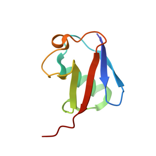
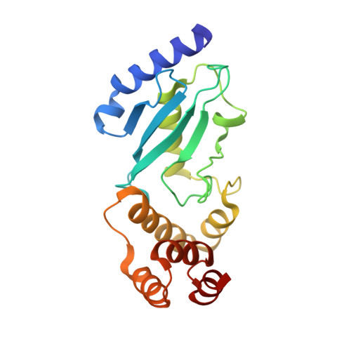
The molecular basis of lysine 48 ubiquitin chain synthesis by Ube2K.

Middleton, A.J.Day, C.L.

(2015) Sci Rep 5: 16793-16793

  • DOI: https://doi.org/10.1038/srep16793
  • Primary Citation of Related Structures:  
    5DFL

  • PubMed Abstract: 

    The post-translational modification of proteins by ubiquitin is central to the regulation of eukaryotic cells. Substrate-bound ubiquitin chains linked by lysine 11 and 48 target proteins to the proteasome for degradation and determine protein abundance in cells, while other ubiquitin chain linkages regulate protein interactions. The specificity of chain-linkage type is usually determined by ubiquitin-conjugating enzymes (E2s). The degradative E2, Ube2K, preferentially catalyses formation of Lys48-linked chains, but like most E2s, the molecular basis for chain formation is not well understood. Here we report the crystal structure of a Ube2K~ubiquitin conjugate and demonstrate that even though it is monomeric, Ube2K can synthesize Lys48-linked ubiquitin chains. Using site-directed mutagenesis and modelling, our studies reveal a molecular understanding of the catalytic complex and identify key features required for synthesis of degradative Lys48-linked chains. The position of the acceptor ubiquitin described here is likely conserved in other E2s that catalyse Lys48-linked ubiquitin chain synthesis.


  • Organizational Affiliation

    Department of Biochemistry, Otago School of Medical Sciences, University of Otago, Dunedin 9054, New Zealand.


Macromolecules
Find similar proteins by:  (by identity cutoff)  |  3D Structure
Entity ID: 1
MoleculeChains Sequence LengthOrganismDetailsImage
UbiquitinA [auth B]76Homo sapiensMutation(s): 0 
Gene Names: UBB
UniProt & NIH Common Fund Data Resources
Find proteins for P0CG47 (Homo sapiens)
Explore P0CG47 
Go to UniProtKB:  P0CG47
PHAROS:  P0CG47
GTEx:  ENSG00000170315 
Entity Groups  
Sequence Clusters30% Identity50% Identity70% Identity90% Identity95% Identity100% Identity
UniProt GroupP0CG47
Sequence Annotations
Expand
  • Reference Sequence
Find similar proteins by:  (by identity cutoff)  |  3D Structure
Entity ID: 2
MoleculeChains Sequence LengthOrganismDetailsImage
Ubiquitin-conjugating enzyme E2 KB [auth A]200Homo sapiensMutation(s): 3 
Gene Names: UBE2KHIP2LIG
EC: 6.3.2.19
UniProt & NIH Common Fund Data Resources
Find proteins for P61086 (Homo sapiens)
Explore P61086 
Go to UniProtKB:  P61086
PHAROS:  P61086
GTEx:  ENSG00000078140 
Entity Groups  
Sequence Clusters30% Identity50% Identity70% Identity90% Identity95% Identity100% Identity
UniProt GroupP61086
Sequence Annotations
Expand
  • Reference Sequence
Experimental Data & Validation

Experimental Data

  • Method: X-RAY DIFFRACTION
  • Resolution: 2.10 Å
  • R-Value Free: 0.235 
  • R-Value Work: 0.195 
  • R-Value Observed: 0.197 
  • Space Group: C 1 2 1
Unit Cell:
Length ( Å )Angle ( ˚ )
a = 146.324α = 90
b = 37.557β = 90.43
c = 61.001γ = 90
Software Package:
Software NamePurpose
Aimlessdata scaling
REFMACrefinement
PDB_EXTRACTdata extraction
XDSdata reduction
PHASERphasing

Structure Validation

View Full Validation Report


Entry History & Funding Information

Deposition Data


Funding OrganizationLocationGrant Number
Marsden Fund, Royal Society of New ZealandNew Zealand--

Revision History  (Full details and data files)

  • Version 1.0: 2015-12-02
    Type: Initial release
  • Version 1.1: 2015-12-09
    Changes: Database references
  • Version 1.2: 2023-09-27
    Changes: Data collection, Database references, Derived calculations, Refinement description