5CS4

Crystal structure of domains AC3-AC5 of yeast acetyl-CoA carboxylase

  • Classification: LIGASE
  • Organism(s): Saccharomyces cerevisiae S288C
  • Expression System: Escherichia coli
  • Mutation(s): No 

  • Deposited: 2015-07-23 Released: 2015-10-28 
  • Deposition Author(s): Wei, J., Tong, L.
  • Funding Organization(s): National Institutes of Health/Office of the Director, National Institutes of Health/National Institute of Diabetes and Digestive and Kidney Disease (NIH/NIDDK)

Experimental Data Snapshot

  • Method: X-RAY DIFFRACTION
  • Resolution: 3.19 Å
  • R-Value Free: 0.287 
  • R-Value Work: 0.234 
  • R-Value Observed: 0.236 

wwPDB Validation   3D Report Full Report


This is version 1.3 of the entry. See complete history


Literature

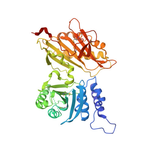
Crystal structure of the 500-kDa yeast acetyl-CoA carboxylase holoenzyme dimer.

Wei, J.Tong, L.

(2015) Nature 526: 723-727

  • DOI: https://doi.org/10.1038/nature15375
  • Primary Citation of Related Structures:  
    5CS0, 5CS4, 5CSA, 5CSK, 5CSL

  • PubMed Abstract: 

    Acetyl-CoA carboxylase (ACC) has crucial roles in fatty acid metabolism and is an attractive target for drug discovery against diabetes, cancer and other diseases. Saccharomyces cerevisiae ACC (ScACC) is crucial for the production of very-long-chain fatty acids and the maintenance of the nuclear envelope. ACC contains biotin carboxylase (BC) and carboxyltransferase (CT) activities, and its biotin is linked covalently to the biotin carboxyl carrier protein (BCCP). Most eukaryotic ACCs are 250-kilodalton (kDa), multi-domain enzymes and function as homodimers and higher oligomers. They contain a unique, 80-kDa central region that shares no homology with other proteins. Although the structures of the BC, CT and BCCP domains and other biotin-dependent carboxylase holoenzymes are known, there is currently no structural information on the ACC holoenzyme. Here we report the crystal structure of the full-length, 500-kDa holoenzyme dimer of ScACC. The structure is remarkably different from that of the other biotin-dependent carboxylases. The central region contains five domains and is important for positioning the BC and CT domains for catalysis. The structure unexpectedly reveals a dimer of the BC domain and extensive conformational differences compared to the structure of the BC domain alone, which is a monomer. These structural changes reveal why the BC domain alone is catalytically inactive and define the molecular mechanism for the inhibition of eukaryotic ACC by the natural product soraphen A and by phosphorylation of a Ser residue just before the BC domain core in mammalian ACC. The BC and CT active sites are separated by 80 Å, and the entire BCCP domain must translocate during catalysis.


  • Organizational Affiliation

    Department of Biological Sciences, Columbia University, New York, New York 10027, USA.


Macromolecules
Find similar proteins by:  (by identity cutoff)  |  3D Structure
Entity ID: 1
MoleculeChains Sequence LengthOrganismDetailsImage
Acetyl-CoA carboxylase
A, B
474Saccharomyces cerevisiae S288CMutation(s): 0 
Gene Names: ACC1ABP2FAS3MTR7YNR016CN3175
EC: 6.4.1.2 (PDB Primary Data), 6.3.4.14 (PDB Primary Data)
UniProt
Find proteins for Q00955 (Saccharomyces cerevisiae (strain ATCC 204508 / S288c))
Explore Q00955 
Go to UniProtKB:  Q00955
Entity Groups  
Sequence Clusters30% Identity50% Identity70% Identity90% Identity95% Identity100% Identity
UniProt GroupQ00955
Sequence Annotations
Expand
  • Reference Sequence
Small Molecules
Modified Residues  1 Unique
IDChains TypeFormula2D DiagramParent
MSE
Query on MSE
A, B
L-PEPTIDE LINKINGC5 H11 N O2 SeMET
Experimental Data & Validation

Experimental Data

  • Method: X-RAY DIFFRACTION
  • Resolution: 3.19 Å
  • R-Value Free: 0.287 
  • R-Value Work: 0.234 
  • R-Value Observed: 0.236 
  • Space Group: P 1 21 1
Unit Cell:
Length ( Å )Angle ( ˚ )
a = 56.849α = 90
b = 93.285β = 100.62
c = 111.144γ = 90
Software Package:
Software NamePurpose
REFMACrefinement
DENZOdata reduction
SCALEPACKdata scaling
SOLVEphasing
RESOLVEphasing
Cootmodel building
PDB_EXTRACTdata extraction

Structure Validation

View Full Validation Report


Entry History & Funding Information

Deposition Data


Funding OrganizationLocationGrant Number
National Institutes of Health/Office of the DirectorUnited StatesS10OD012018
National Institutes of Health/National Institute of Diabetes and Digestive and Kidney Disease (NIH/NIDDK)United StatesR01DK067238

Revision History  (Full details and data files)

  • Version 1.0: 2015-10-28
    Type: Initial release
  • Version 1.1: 2015-11-11
    Changes: Database references
  • Version 1.2: 2017-09-20
    Changes: Author supporting evidence, Database references, Derived calculations
  • Version 1.3: 2019-12-25
    Changes: Author supporting evidence