5BNB

Crystal structure of a Ube2S-ubiquitin conjugate


Experimental Data Snapshot

  • Method: X-RAY DIFFRACTION
  • Resolution: 2.49 Å
  • R-Value Free: 0.304 
  • R-Value Work: 0.243 
  • R-Value Observed: 0.246 

wwPDB Validation   3D Report Full Report


This is version 1.2 of the entry. See complete history


Literature

Crystal Structure of a Ube2S-Ubiquitin Conjugate.

Lorenz, S.Bhattacharyya, M.Feiler, C.Rape, M.Kuriyan, J.

(2016) PLoS One 11: e0147550-e0147550

  • DOI: https://doi.org/10.1371/journal.pone.0147550
  • Primary Citation of Related Structures:  
    5BNB

  • PubMed Abstract: 

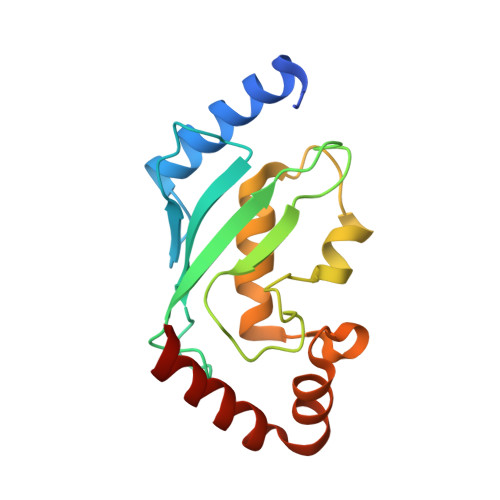
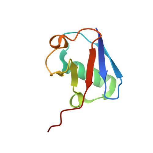
    Protein ubiquitination occurs through the sequential formation and reorganization of specific protein-protein interfaces. Ubiquitin-conjugating (E2) enzymes, such as Ube2S, catalyze the formation of an isopeptide linkage between the C-terminus of a "donor" ubiquitin and a primary amino group of an "acceptor" ubiquitin molecule. This reaction involves an intermediate, in which the C-terminus of the donor ubiquitin is thioester-bound to the active site cysteine of the E2 and a functionally important interface is formed between the two proteins. A docked model of a Ube2S-donor ubiquitin complex was generated previously, based on chemical shift mapping by NMR, and predicted contacts were validated in functional studies. We now present the crystal structure of a covalent Ube2S-ubiquitin complex. The structure contains an interface between Ube2S and ubiquitin in trans that resembles the earlier model in general terms, but differs in detail. The crystallographic interface is more hydrophobic than the earlier model and is stable in molecular dynamics (MD) simulations. Remarkably, the docked Ube2S-donor complex converges readily to the configuration seen in the crystal structure in 3 out of 8 MD trajectories. Since the crystallographic interface is fully consistent with mutational effects, this indicates that the structure provides an energetically favorable representation of the functionally critical Ube2S-donor interface.


  • Organizational Affiliation

    California Institute for Quantitative Biosciences, University of California, Berkeley, California, United States of America.


Macromolecules
Find similar proteins by:  (by identity cutoff)  |  3D Structure
Entity ID: 1
MoleculeChains Sequence LengthOrganismDetailsImage
Ubiquitin-conjugating enzyme E2 SA,
B,
C,
H [auth D]
156Homo sapiensMutation(s): 1 
Gene Names: UBE2SE2EPFOK/SW-cl.73
EC: 6.3.2.19
UniProt & NIH Common Fund Data Resources
Find proteins for Q16763 (Homo sapiens)
Explore Q16763 
Go to UniProtKB:  Q16763
PHAROS:  Q16763
GTEx:  ENSG00000108106 
Entity Groups  
Sequence Clusters30% Identity50% Identity70% Identity90% Identity95% Identity100% Identity
UniProt GroupQ16763
Sequence Annotations
Expand
  • Reference Sequence
Find similar proteins by:  (by identity cutoff)  |  3D Structure
Entity ID: 2
MoleculeChains Sequence LengthOrganismDetailsImage
Polyubiquitin-BD [auth E],
E [auth F],
F [auth G],
G [auth I]
76Homo sapiensMutation(s): 1 
Gene Names: UBB
UniProt & NIH Common Fund Data Resources
Find proteins for P0CG47 (Homo sapiens)
Explore P0CG47 
Go to UniProtKB:  P0CG47
PHAROS:  P0CG47
GTEx:  ENSG00000170315 
Entity Groups  
Sequence Clusters30% Identity50% Identity70% Identity90% Identity95% Identity100% Identity
UniProt GroupP0CG47
Sequence Annotations
Expand
  • Reference Sequence
Experimental Data & Validation

Experimental Data

  • Method: X-RAY DIFFRACTION
  • Resolution: 2.49 Å
  • R-Value Free: 0.304 
  • R-Value Work: 0.243 
  • R-Value Observed: 0.246 
  • Space Group: P 1 21 1
Unit Cell:
Length ( Å )Angle ( ˚ )
a = 94.906α = 90
b = 45.854β = 93.14
c = 110.576γ = 90
Software Package:
Software NamePurpose
PHENIXrefinement
XDSdata reduction
Aimlessdata scaling
PHASERphasing

Structure Validation

View Full Validation Report


Entry History & Funding Information

Deposition Data


Funding OrganizationLocationGrant Number
The Leukemia and Lymphoma SocietyUnited States5509-11
German Research FoundationGermanyLO 2003/1-1

Revision History  (Full details and data files)

  • Version 1.0: 2016-02-17
    Type: Initial release
  • Version 1.1: 2017-09-06
    Changes: Author supporting evidence
  • Version 1.2: 2024-01-10
    Changes: Data collection, Database references, Refinement description