


Crystal Structure of a Phosphoserine/phosphohydroxythreonine Aminotransferase (PSAT) from Pseudomonas aeruginosa with cofactor Pyridoxal Phosphate and bound Glutamate
SSGCID, Dranow, D.M., Abendroth, J., Lorimer, D., Edwards, T.E.To be published.
Experimental Data Snapshot
wwPDB Validation 3D Report Full Report

Entity ID: 1 | |||||
|---|---|---|---|---|---|
| Molecule | Chains | Sequence Length | Organism | Details | Image |
| Phosphoserine aminotransferase | 369 | Pseudomonas aeruginosa PAO1 | Mutation(s): 0 Gene Names: serC, PA3167 EC: 2.6.1.52 | 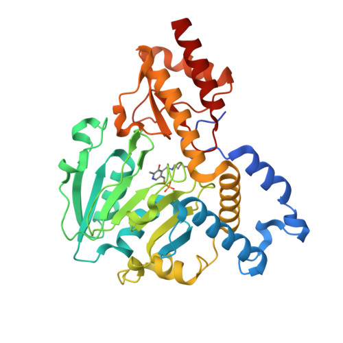 | |
UniProt | |||||
Find proteins for Q9HZ66 (Pseudomonas aeruginosa (strain ATCC 15692 / DSM 22644 / CIP 104116 / JCM 14847 / LMG 12228 / 1C / PRS 101 / PAO1)) Explore Q9HZ66 Go to UniProtKB: Q9HZ66 | |||||
Entity Groups | |||||
| Sequence Clusters | 30% Identity50% Identity70% Identity90% Identity95% Identity100% Identity | ||||
| UniProt Group | Q9HZ66 | ||||
Sequence AnnotationsExpand | |||||
| |||||
| Ligands 2 Unique | |||||
|---|---|---|---|---|---|
| ID | Chains | Name / Formula / InChI Key | 2D Diagram | 3D Interactions | |
| GLU Query on GLU | F [auth B] | GLUTAMIC ACID C5 H9 N O4 WHUUTDBJXJRKMK-VKHMYHEASA-N |  | ||
| FMT Query on FMT | C [auth A], D [auth A], E [auth B] | FORMIC ACID C H2 O2 BDAGIHXWWSANSR-UHFFFAOYSA-N |  | ||
| Modified Residues 1 Unique | |||||
|---|---|---|---|---|---|
| ID | Chains | Type | Formula | 2D Diagram | Parent |
| LLP Query on LLP | A, B | L-PEPTIDE LINKING | C14 H22 N3 O7 P |  | LYS |
| Length ( Å ) | Angle ( ˚ ) |
|---|---|
| a = 72.22 | α = 90 |
| b = 72.22 | β = 90 |
| c = 268.48 | γ = 120 |
| Software Name | Purpose |
|---|---|
| XSCALE | data scaling |
| PHENIX | refinement |
| PDB_EXTRACT | data extraction |
| ARP | model building |
RCSB PDB (citation) is hosted by
RCSB PDB is a member of the

