4UUK

Human dynamin 1 K44A superconstricted polymer stabilized with GTP strand 2


Experimental Data Snapshot

  • Method: ELECTRON MICROSCOPY
  • Resolution: 12.5 Å
  • Aggregation State: FILAMENT 
  • Reconstruction Method: HELICAL 

wwPDB Validation   3D Report Full Report


This is version 1.1 of the entry. See complete history


Literature

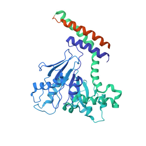
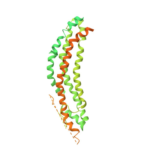
A Dynamin Mutant Defines a Superconstricted Prefission State.

Sundborger, A.C.Fang, S.Heymann, J.A.Ray, P.Chappie, J.S.Hinshaw, J.E.

(2014) Cell Rep 8: 734

  • DOI: https://doi.org/10.1016/j.celrep.2014.06.054
  • Primary Citation of Related Structures:  
    4UUD, 4UUK

  • PubMed Abstract: 

    Dynamin is a 100 kDa GTPase that organizes into helical assemblies at the base of nascent clathrin-coated vesicles. Formation of these oligomers stimulates the intrinsic GTPase activity of dynamin, which is necessary for efficient membrane fission during endocytosis. Recent evidence suggests that the transition state of dynamin's GTP hydrolysis reaction serves as a key determinant of productive fission. Here, we present the structure of a transition-state-defective dynamin mutant K44A trapped in a prefission state at 12.5 Å resolution. This structure constricts to 3.7 nm, reaching the theoretical limit required for spontaneous membrane fission. Computational docking indicates that the ground-state conformation of the dynamin polymer is sufficient to achieve this superconstricted prefission state and reveals how a two-start helical symmetry promotes the most efficient packing of dynamin tetramers around the membrane neck. These data suggest a model for the assembly and regulation of the minimal dynamin fission machine.


  • Organizational Affiliation

    Laboratory of Cell and Molecular Biology, National Institute of Diabetes and Digestive and Kidney Diseases, National Institutes of Health, Bethesda, MD 20892, USA.


Macromolecules
Find similar proteins by:  (by identity cutoff)  |  3D Structure
Entity ID: 1
MoleculeChains Sequence LengthOrganismDetailsImage
DYNAMIN-1
A, D, G, K
864Homo sapiensMutation(s): 1 
EC: 3.6.5.5
UniProt & NIH Common Fund Data Resources
Find proteins for Q05193 (Homo sapiens)
Explore Q05193 
Go to UniProtKB:  Q05193
PHAROS:  Q05193
GTEx:  ENSG00000106976 
Entity Groups  
Sequence Clusters30% Identity50% Identity70% Identity90% Identity95% Identity100% Identity
UniProt GroupQ05193
Sequence Annotations
Expand
  • Reference Sequence
Find similar proteins by:  (by identity cutoff)  |  3D Structure
Entity ID: 2
MoleculeChains Sequence LengthOrganismDetailsImage
DYNAMIN-1
B, C, E, F, H
B, C, E, F, H, I, J, L
864Homo sapiensMutation(s): 0 
EC: 3.6.5.5
UniProt & NIH Common Fund Data Resources
Find proteins for Q05193 (Homo sapiens)
Explore Q05193 
Go to UniProtKB:  Q05193
PHAROS:  Q05193
GTEx:  ENSG00000106976 
Entity Groups  
Sequence Clusters30% Identity50% Identity70% Identity90% Identity95% Identity100% Identity
UniProt GroupQ05193
Sequence Annotations
Expand
  • Reference Sequence
Experimental Data & Validation

Experimental Data

  • Method: ELECTRON MICROSCOPY
  • Resolution: 12.5 Å
  • Aggregation State: FILAMENT 
  • Reconstruction Method: HELICAL 
EM Software:
TaskSoftware PackageVersion
RECONSTRUCTIONSPIDER

Structure Validation

View Full Validation Report


Entry History 

Deposition Data

Revision History  (Full details and data files)

  • Version 1.0: 2014-08-27
    Type: Initial release
  • Version 1.1: 2017-08-30
    Changes: Data collection