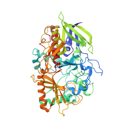
High-resolution structures of cholesterol oxidase in the reduced state provide insights into redox stabilization.
Golden, E., Karton, A., Vrielink, A.(2014) Acta Crystallogr D Biol Crystallogr 70: 3155-3166
- PubMed: 25478834
- DOI: https://doi.org/10.1107/S139900471402286X
- Primary Citation of Related Structures:
4U2L, 4U2S, 4U2T - PubMed Abstract:
Cholesterol oxidase (CO) is a flavoenzyme that catalyzes the oxidation and isomerization of cholesterol to cholest-4-en-3-one. The reductive half reaction occurs via a hydride transfer from the substrate to the FAD cofactor. The structures of CO reduced with dithionite under aerobic conditions and in the presence of the substrate 2-propanol under both aerobic and anaerobic conditions are presented. The 1.32 Å resolution structure of the dithionite-reduced enzyme reveals a sulfite molecule covalently bound to the FAD cofactor. The isoalloxazine ring system displays a bent structure relative to that of the oxidized enzyme, and alternate conformations of a triad of aromatic residues near to the cofactor are evident. A 1.12 Å resolution anaerobically trapped reduced enzyme structure in the presence of 2-propanol does not show a similar bending of the flavin ring system, but does show alternate conformations of the aromatic triad. Additionally, a significant difference electron-density peak is observed within a covalent-bond distance of N5 of the flavin moiety, suggesting that a hydride-transfer event has occurred as a result of substrate oxidation trapping the flavin in the electron-rich reduced state. The hydride transfer generates a tetrahedral geometry about the flavin N5 atom. High-level density-functional theory calculations were performed to correlate the crystallographic findings with the energetics of this unusual arrangement of the flavin moiety. These calculations suggest that strong hydrogen-bond interactions between Gly120 and the flavin N5 centre may play an important role in these structural features.
Organizational Affiliation:
School of Chemistry and Biochemistry, University of Western Australia, Crawley, Western Australia 6009, Australia.


