4RSG

Neutron crystal structure of Ras bound to the GTP analogue GppNHp


Experimental Data Snapshot

  • Method: NEUTRON DIFFRACTION
  • Resolution: 1.91 Å
  • R-Value Free: 0.287 
  • R-Value Work: 0.249 
  • R-Value Observed: 0.253 

wwPDB Validation   3D Report Full Report

Currently 4RSG does not have a validation slider image.


This is version 1.5 of the entry. See complete history


Literature

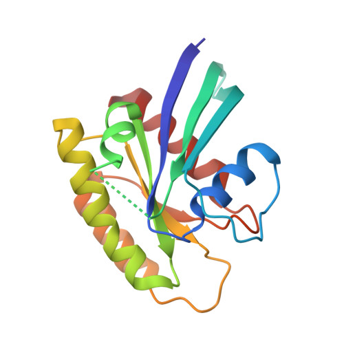
Neutron Crystal Structure of RAS GTPase Puts in Question the Protonation State of the GTP gamma-Phosphate.

Knihtila, R.Holzapfel, G.Weiss, K.Meilleur, F.Mattos, C.

(2015) J Biol Chem 290: 31025-31036

  • DOI: https://doi.org/10.1074/jbc.M115.679860
  • Primary Citation of Related Structures:  
    4RSG

  • PubMed Abstract: 

    RAS GTPase is a prototype for nucleotide-binding proteins that function by cycling between GTP and GDP, with hydrogen atoms playing an important role in the GTP hydrolysis mechanism. It is one of the most well studied proteins in the superfamily of small GTPases, which has representatives in a wide range of cellular functions. These proteins share a GTP-binding pocket with highly conserved motifs that promote hydrolysis to GDP. The neutron crystal structure of RAS presented here strongly supports a protonated γ-phosphate at physiological pH. This counters the notion that the phosphate groups of GTP are fully deprotonated at the start of the hydrolysis reaction, which has colored the interpretation of experimental and computational data in studies of the hydrolysis mechanism. The neutron crystal structure presented here puts in question our understanding of the pre-catalytic state associated with the hydrolysis reaction central to the function of RAS and other GTPases.


  • Organizational Affiliation

    From the Department of Chemistry and Chemical Biology, Northeastern University, Boston, Massachusetts 02115.


Macromolecules
Find similar proteins by:  (by identity cutoff)  |  3D Structure
Entity ID: 1
MoleculeChains Sequence LengthOrganismDetailsImage
GTPase HRas166Homo sapiensMutation(s): 0 
Gene Names: HRASHRAS1
UniProt & NIH Common Fund Data Resources
Find proteins for P01112 (Homo sapiens)
Explore P01112 
Go to UniProtKB:  P01112
PHAROS:  P01112
GTEx:  ENSG00000174775 
Entity Groups  
Sequence Clusters30% Identity50% Identity70% Identity90% Identity95% Identity100% Identity
UniProt GroupP01112
Sequence Annotations
Expand
  • Reference Sequence
Small Molecules
Ligands 2 Unique
IDChains Name / Formula / InChI Key2D Diagram3D Interactions
GNP
Query on GNP

Download Ideal Coordinates CCD File 
B [auth A]PHOSPHOAMINOPHOSPHONIC ACID-GUANYLATE ESTER
C10 H17 N6 O13 P3
UQABYHGXWYXDTK-UUOKFMHZSA-N
MG
Query on MG

Download Ideal Coordinates CCD File 
C [auth A]MAGNESIUM ION
Mg
JLVVSXFLKOJNIY-UHFFFAOYSA-N
Experimental Data & Validation

Experimental Data

  • Method: NEUTRON DIFFRACTION
  • Resolution: 1.91 Å
  • R-Value Free: 0.287 
  • R-Value Work: 0.249 
  • R-Value Observed: 0.253 
  • Space Group: P 32 2 1
Unit Cell:
Length ( Å )Angle ( ˚ )
a = 40.084α = 90
b = 40.084β = 90
c = 160.785γ = 120
Software Package:
Software NamePurpose
SCALAdata scaling
PHENIXrefinement
PDB_EXTRACTdata extraction
Quasi-Lauedata collection
LAUEGENdata reduction
PHENIXphasing

Structure Validation

View Full Validation Report

Currently 4RSG does not have a validation slider image.


Entry History 

Deposition Data

Revision History  (Full details and data files)

  • Version 1.0: 2015-11-04
    Type: Initial release
  • Version 1.1: 2015-11-18
    Changes: Database references
  • Version 1.2: 2016-01-13
    Changes: Database references
  • Version 1.3: 2018-04-25
    Changes: Data collection
  • Version 1.4: 2021-06-30
    Changes: Data collection, Derived calculations
  • Version 1.5: 2024-02-28
    Changes: Data collection, Database references