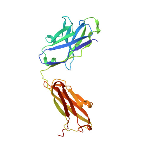
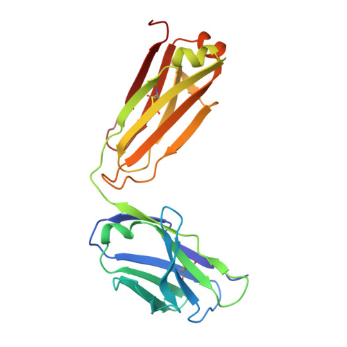
Stabilizing a flexible interdomain hinge region harboring the SMB binding site drives uPAR into its closed conformation.
Zhao, B., Gandhi, S., Yuan, C., Luo, Z., Li, R., Gardsvoll, H., de Lorenzi, V., Sidenius, N., Huang, M., Ploug, M.(2015) J Mol Biol 427: 1389-1403
- PubMed: 25659907
- DOI: https://doi.org/10.1016/j.jmb.2015.01.022
- Primary Citation of Related Structures:
4QTH, 4QTI - PubMed Abstract:
The urokinase-type plasminogen activator receptor (uPAR) is a multidomain glycolipid-anchored membrane protein, which facilitates extracellular matrix remodeling by focalizing plasminogen activation to cell surfaces via its high-affinity interaction with uPA. The modular assembly of its three LU (Ly6/uPAR-like) domains is inherently flexible and binding of uPA drives uPAR into its closed conformation, which presents the higher-affinity state for vitronectin thus providing an allosteric regulatory mechanism. Using a new class of epitope-mapped anti-uPAR monoclonal antibodies (mAbs), we now demonstrate that the reciprocal stabilization is indeed also possible. By surface plasmon resonance studies, we show that these mAbs and vitronectin have overlapping binding sites on uPAR and that they share Arg91 as hotspot residue in their binding interfaces. The crystal structure solved for one of these uPAR·mAb complexes at 3.0Å clearly shows that this mAb preselects the closed uPAR conformation with an empty but correctly assembled large hydrophobic binding cavity for uPA. Accordingly, these mAbs inhibit the uPAR-dependent lamellipodia formation and migration on vitronectin-coated matrices irrespective of the conformational status of uPAR and its occupancy with uPA. This is the first study to the best of our knowledge, showing that the dynamic assembly of the three LU domains in uPARwt can be driven toward the closed form by an external ligand, which is not engaging the hydrophobic uPA binding cavity. As this binding interface is also exploited by the somatomedin B domain of vitronectin, therefore, this relationship should be taken into consideration when exploring uPAR-dependent cell adhesion and migration in vitronectin-rich environments.
Organizational Affiliation:
State Key Laboratory of Structural Chemistry, Fujian Institute of Research on the Structure of Matter, Chinese Academy of Sciences, Fuzhou, Fujian 350002, China; Danish-Chinese Centre for Proteases and Cancer.