4OXX

Crystal Structure of Cindoxin, Surface Entropy reduction Mutant


Experimental Data Snapshot

  • Method: X-RAY DIFFRACTION
  • Resolution: 1.21 Å
  • R-Value Free: 0.160 
  • R-Value Work: 0.141 
  • R-Value Observed: 0.142 

wwPDB Validation   3D Report Full Report


Ligand Structure Quality Assessment 


This is version 1.4 of the entry. See complete history


Literature

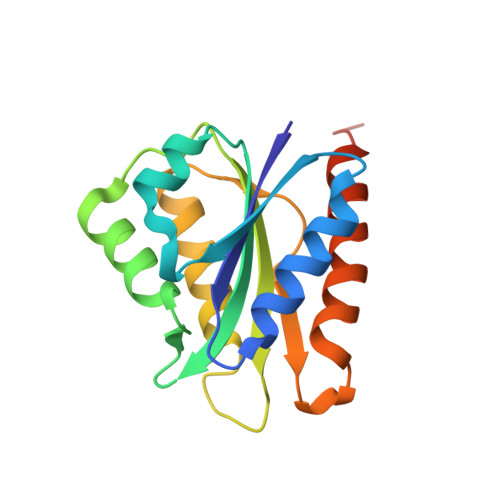
Crystal structure of cindoxin, the P450cin redox partner.

Madrona, Y.Hollingsworth, S.A.Tripathi, S.Fields, J.B.Rwigema, J.C.Tobias, D.J.Poulos, T.L.

(2014) Biochemistry 53: 1435-1446

  • DOI: https://doi.org/10.1021/bi500010m
  • Primary Citation of Related Structures:  
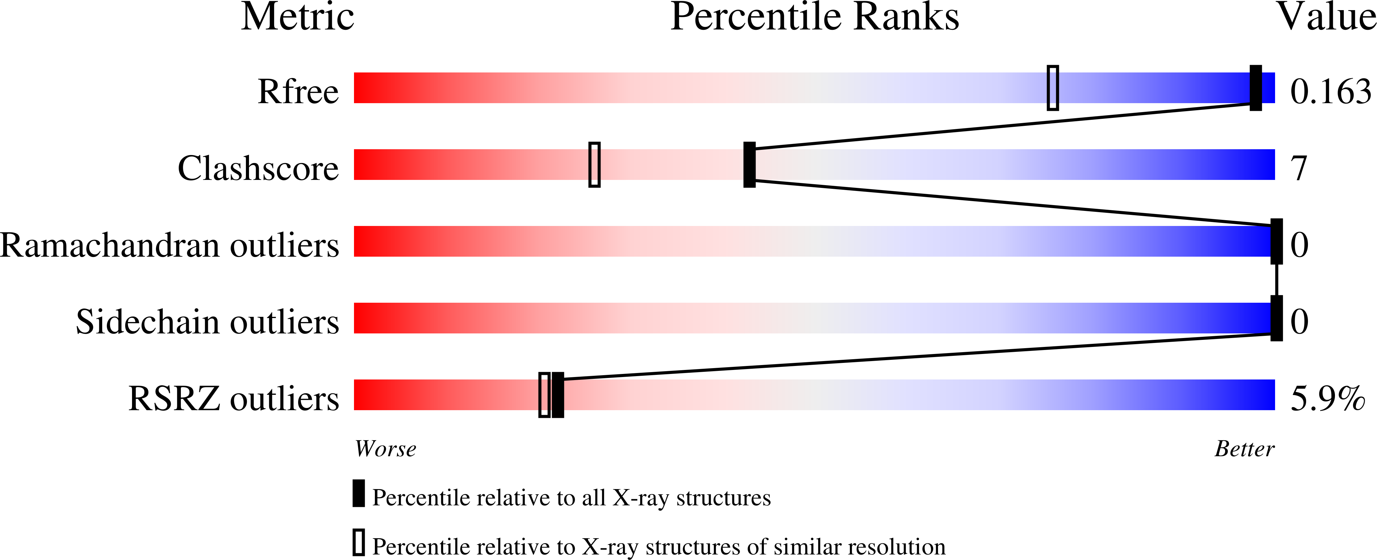
    4OXX

  • PubMed Abstract: 

    The crystal structure of the flavin mononucleotide (FMN)-containing redox partner to P450cin, cindoxin (Cdx), has been determined to 1.3 Å resolution. The overall structure is similar to that of the FMN domain of human cytochrome P450 reductase. A Brownian dynamics-molecular dynamics docking method was used to produce a model of Cdx with its redox partner, P450cin. This Cdx-P450cin model highlights the potential importance of Cdx Tyr96 in bridging the FMN and heme cofactors as well P450cin Arg102 and Arg346. Each of the single-site Ala mutants exhibits ~10% of the wild-type activity, thus demonstrating the importance of these residues for binding and/or electron transfer. In the well-studied P450cam system, redox partner binding stabilizes the open low-spin conformation of P450cam and greatly decreases the stability of the oxy complex. In sharp contrast, Cdx does not shift P450cin to a low-spin state, although the stability of oxy-P450cin is decreased 10-fold in the presence of Cdx. This indicates that Cdx may have a modest effect on the open-closed equilibrium in P450cin compared to that in P450cam. It has been postulated that part of the effector role of Pdx on P450cam is to promote a significant structural change that makes available a proton relay network involving Asp251 required for O2 activation. The structure around the corresponding Asp in P450cin, Asp241, provides a possible structural reason for why P450cin is less dependent on its redox partner for functionally important structural changes.


  • Organizational Affiliation

    Departments of †Molecular Biology and Biochemistry, ‡Chemistry, and §Pharmaceutical Sciences, University of California , Irvine, California 92697-3900, United States.


Macromolecules
Find similar proteins by:  (by identity cutoff)  |  3D Structure
Entity ID: 1
MoleculeChains Sequence LengthOrganismDetailsImage
Cindoxin162Citrobacter braakiiMutation(s): 3 
Gene Names: cinC
UniProt
Find proteins for Q8VQF4 (Citrobacter braakii)
Explore Q8VQF4 
Go to UniProtKB:  Q8VQF4
Entity Groups  
Sequence Clusters30% Identity50% Identity70% Identity90% Identity95% Identity100% Identity
UniProt GroupQ8VQF4
Sequence Annotations
Expand
  • Reference Sequence
Small Molecules
Ligands 1 Unique
IDChains Name / Formula / InChI Key2D Diagram3D Interactions
FMN
Query on FMN

Download Ideal Coordinates CCD File 
B [auth A]FLAVIN MONONUCLEOTIDE
C17 H21 N4 O9 P
FVTCRASFADXXNN-SCRDCRAPSA-N
Experimental Data & Validation

Experimental Data

  • Method: X-RAY DIFFRACTION
  • Resolution: 1.21 Å
  • R-Value Free: 0.160 
  • R-Value Work: 0.141 
  • R-Value Observed: 0.142 
  • Space Group: P 32 2 1
Unit Cell:
Length ( Å )Angle ( ˚ )
a = 82.109α = 90
b = 82.109β = 90
c = 37.042γ = 120
Software Package:
Software NamePurpose
PHENIXrefinement

Structure Validation

View Full Validation Report


Ligand Structure Quality Assessment 


Entry History & Funding Information

Deposition Data


Funding OrganizationLocationGrant Number
National Institutes of Health/National Institute of General Medical Sciences (NIH/NIGMS)United StatesGM33688

Revision History  (Full details and data files)

  • Version 1.0: 2014-04-16
    Type: Initial release
  • Version 1.1: 2014-10-01
    Changes: Database references
  • Version 1.2: 2017-09-20
    Changes: Advisory, Author supporting evidence, Database references, Derived calculations, Other, Source and taxonomy
  • Version 1.3: 2019-12-25
    Changes: Author supporting evidence
  • Version 1.4: 2023-12-27
    Changes: Data collection, Database references, Refinement description