Crystal structure and immunological properties of the first annexin from Schistosoma mansoni: insights into the structural integrity of the schistosomal tegument.
Leow, C.Y., Willis, C., Osman, A., Mason, L., Simon, A., Smith, B.J., Gasser, R.B., Jones, M.K., Hofmann, A.(2014) FEBS J 281: 1209-1225
- PubMed: 24428567
- DOI: https://doi.org/10.1111/febs.12700
- Primary Citation of Related Structures:
4MDU, 4MDV - PubMed Abstract:
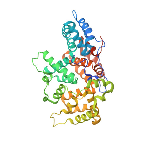
Schistosomiasis is a major parasitic disease of humans, second only to malaria in its global impact. The disease is caused by digenean trematodes that infest the vasculature of their human hosts. These flukes are limited externally by a body wall composed of a syncytial epithelium, the apical surface membrane of which is a parasitism-adapted dual membrane complex. Annexins are thought to be of integral importance for the stability of this apical membrane system. Here, we present the first structural and immunobiochemical characterization of an annexin from Schistosoma mansoni. The crystal structure of annexin B22 confirms the presence of the previously predicted α-helical segment in the II/III linker and reveals a covalently linked head-to-head dimer. From the calcium-bound crystal structure of this protein, canonical type II, type III and B site positions are occupied, and a novel binding site has been identified. The dimer arrangement observed in the crystal structure suggests the presence of two prominent features, a potential non-canonical membrane binding site and a potential binding groove opposite to the former. Results from transcriptional profiling during development show that annexin B22 expression is correlated with life stages of the parasite that possess the syncytial tegument layer, and ultrastructural localization by immuno-electron microscopy confirms the occurrence of annexins in the tegument of S. mansoni. Data from membrane binding and aggregation assays indicate the presence of differential molecular mechanisms and support the hypothesis of annexin B22 providing structural integrity in the tegument.
Organizational Affiliation:
School of Veterinary Science, University of Queensland, Gatton, Australia; Queensland Institute of Medical Research, Herston, Australia; Institute for Research in Molecular Medicine, Universiti Sains Malaysia, Penang, Malaysia.