


Crystal structure of ryanodine receptor disease mutant
Kimlicka, L., Tung, C.C., Carlsson, A.C., Lobo, P.A., Van Petegem, F.To be published.
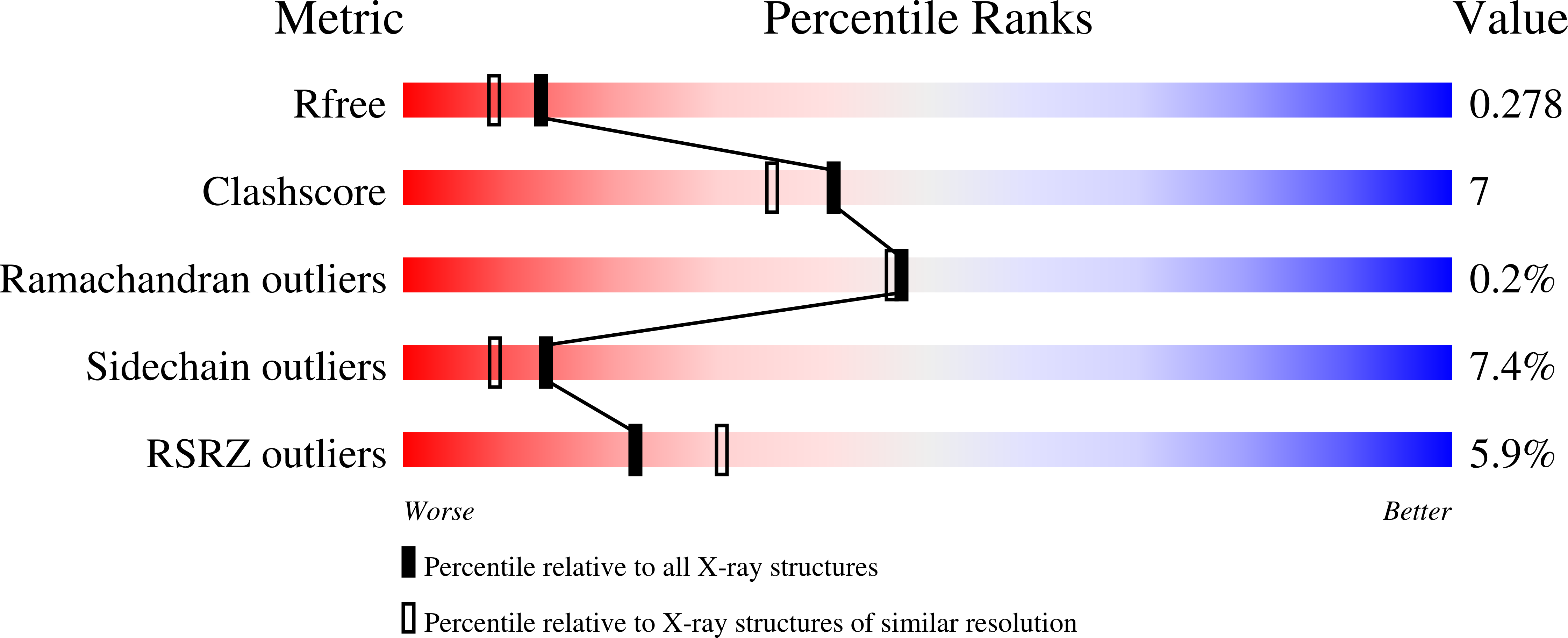
Experimental Data Snapshot
wwPDB Validation 3D Report Full Report
Entity ID: 1 | |||||
|---|---|---|---|---|---|
| Molecule | Chains | Sequence Length | Organism | Details | Image |
| Ryanodine receptor 2 | 547 | Mus musculus | Mutation(s): 1 Gene Names: Ryr2 | 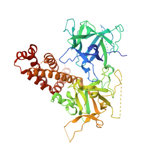 | |
UniProt & NIH Common Fund Data Resources | |||||
Find proteins for E9Q401 (Mus musculus) Explore E9Q401 Go to UniProtKB: E9Q401 | |||||
IMPC: MGI:99685 | |||||
Entity Groups | |||||
| Sequence Clusters | 30% Identity50% Identity70% Identity90% Identity95% Identity100% Identity | ||||
| UniProt Group | E9Q401 | ||||
Sequence AnnotationsExpand | |||||
| |||||
| Ligands 1 Unique | |||||
|---|---|---|---|---|---|
| ID | Chains | Name / Formula / InChI Key | 2D Diagram | 3D Interactions | |
| GOL Query on GOL | B [auth A] | GLYCEROL C3 H8 O3 PEDCQBHIVMGVHV-UHFFFAOYSA-N |  | ||
| Length ( Å ) | Angle ( ˚ ) |
|---|---|
| a = 78.05 | α = 90 |
| b = 78.05 | β = 90 |
| c = 248.1 | γ = 90 |
| Software Name | Purpose |
|---|---|
| HKL-2000 | data collection |
| PHASER | phasing |
| REFMAC | refinement |
| XDS | data reduction |
| XDS | data scaling |
RCSB PDB (citation) is hosted by
RCSB PDB is a member of the

