4KBO

Crystal structure of the human Mortalin (GRP75) ATPase domain in the apo form


Experimental Data Snapshot

  • Method: X-RAY DIFFRACTION
  • Resolution: 2.80 Å
  • R-Value Free: 0.271 
  • R-Value Work: 0.218 
  • R-Value Observed: 0.221 

wwPDB Validation   3D Report Full Report


This is version 1.5 of the entry. See complete history


Literature

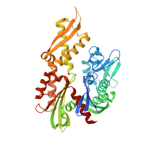
Crystal structure of the nucleotide-binding domain of mortalin, the mitochondrial Hsp70 chaperone.

Amick, J.Schlanger, S.E.Wachnowsky, C.Moseng, M.A.Emerson, C.C.Dare, M.Luo, W.I.Ithychanda, S.S.Nix, J.C.Cowan, J.A.Page, R.C.Misra, S.

(2014) Protein Sci 23: 833-842

  • DOI: https://doi.org/10.1002/pro.2466
  • Primary Citation of Related Structures:  
    4KBO

  • PubMed Abstract: 

    Mortalin, a member of the Hsp70-family of molecular chaperones, functions in a variety of processes including mitochondrial protein import and quality control, Fe-S cluster protein biogenesis, mitochondrial homeostasis, and regulation of p53. Mortalin is implicated in regulation of apoptosis, cell stress response, neurodegeneration, and cancer and is a target of the antitumor compound MKT-077. Like other Hsp70-family members, Mortalin consists of a nucleotide-binding domain (NBD) and a substrate-binding domain. We determined the crystal structure of the NBD of human Mortalin at 2.8 Å resolution. Although the Mortalin nucleotide-binding pocket is highly conserved relative to other Hsp70 family members, we find that its nucleotide affinity is weaker than that of Hsc70. A Parkinson's disease-associated mutation is located on the Mortalin-NBD surface and may contribute to Mortalin aggregation. We present structure-based models for how the Mortalin-NBD may interact with the nucleotide exchange factor GrpEL1, with p53, and with MKT-077. Our structure may contribute to the understanding of disease-associated Mortalin mutations and to improved Mortalin-targeting antitumor compounds.


  • Organizational Affiliation

    Department of Molecular Cardiology, The Cleveland Clinic, Cleveland, Ohio, 44195.


Macromolecules
Find similar proteins by:  (by identity cutoff)  |  3D Structure
Entity ID: 1
MoleculeChains Sequence LengthOrganismDetailsImage
Stress-70 protein, mitochondrial385Homo sapiensMutation(s): 0 
Gene Names: GRP75HSPA9HSPA9B
UniProt & NIH Common Fund Data Resources
Find proteins for P38646 (Homo sapiens)
Explore P38646 
Go to UniProtKB:  P38646
PHAROS:  P38646
GTEx:  ENSG00000113013 
Entity Groups  
Sequence Clusters30% Identity50% Identity70% Identity90% Identity95% Identity100% Identity
UniProt GroupP38646
Sequence Annotations
Expand
  • Reference Sequence
Small Molecules
Ligands 1 Unique
IDChains Name / Formula / InChI Key2D Diagram3D Interactions
NA
Query on NA

Download Ideal Coordinates CCD File 
B [auth A]SODIUM ION
Na
FKNQFGJONOIPTF-UHFFFAOYSA-N
Experimental Data & Validation

Experimental Data

  • Method: X-RAY DIFFRACTION
  • Resolution: 2.80 Å
  • R-Value Free: 0.271 
  • R-Value Work: 0.218 
  • R-Value Observed: 0.221 
  • Space Group: P 21 21 21
Unit Cell:
Length ( Å )Angle ( ˚ )
a = 51.197α = 90
b = 67.812β = 90
c = 120.769γ = 90
Software Package:
Software NamePurpose
ADSCdata collection
PHENIXmodel building
PHENIXrefinement
HKL-3000data reduction
SCALAdata scaling
PHENIXphasing

Structure Validation

View Full Validation Report


Entry History 

Deposition Data

Revision History  (Full details and data files)

  • Version 1.0: 2014-04-02
    Type: Initial release
  • Version 1.1: 2014-04-23
    Changes: Database references
  • Version 1.2: 2014-05-07
    Changes: Other
  • Version 1.3: 2014-05-21
    Changes: Database references
  • Version 1.4: 2014-06-04
    Changes: Database references
  • Version 1.5: 2023-09-20
    Changes: Data collection, Database references, Derived calculations, Refinement description