4FNI

Crystal structure of IsdI-W66Y in complex with heme and cyanide


Experimental Data Snapshot

  • Method: X-RAY DIFFRACTION
  • Resolution: 1.80 Å
  • R-Value Free: 0.238 
  • R-Value Work: 0.197 
  • R-Value Observed: 0.199 

wwPDB Validation   3D Report Full Report


Ligand Structure Quality Assessment 


This is version 1.3 of the entry. See complete history


Literature

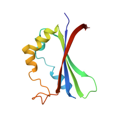
Inactivation of the heme degrading enzyme IsdI by an active site substitution that diminishes heme ruffling.

Ukpabi, G.Takayama, S.J.Mauk, A.G.Murphy, M.E.

(2012) J Biol Chem 287: 34179-34188

  • DOI: https://doi.org/10.1074/jbc.M112.393249
  • Primary Citation of Related Structures:  
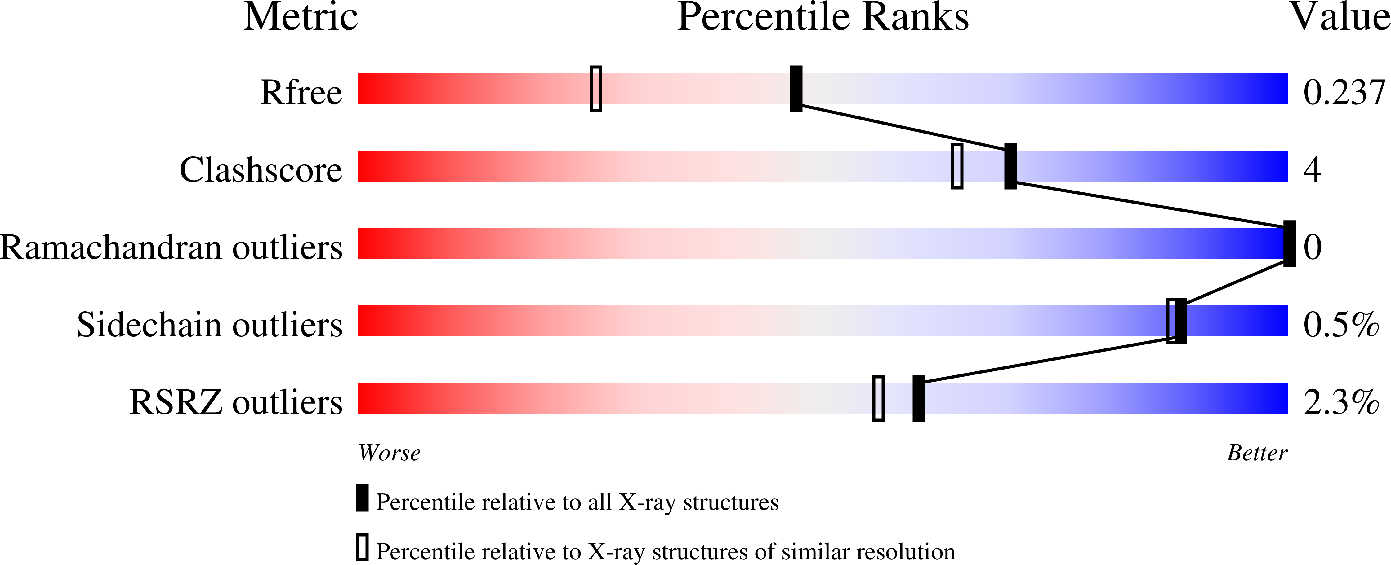
    4FNH, 4FNI

  • PubMed Abstract: 

    IsdG and IsdI are paralogous heme degrading enzymes from the bacterium Staphylococcus aureus. Heme bound by these enzymes is extensively ruffled such that the meso-carbons at the sites of oxidation are distorted toward bound oxygen. In contrast, the canonical heme oxygenase family degrades heme that is bound with minimal distortion. Trp-66 is a conserved heme pocket residue in IsdI implicated in heme ruffling. IsdI variants with Trp-66 replaced with residues having less bulky aromatic and alkyl side chains were characterized with respect to catalytic activity, heme ruffling, and electrochemical properties. The heme degradation activity of the W66Y and W66F variants was approximately half that of the wild-type enzyme, whereas the W66L and W66A variants were inactive. A crystal structure and NMR spectroscopic analysis of the W66Y variant reveals that heme binds to this enzyme with less heme ruffling than observed for wild-type IsdI. The reduction potential of this variant (-96 ± 7 mV versus standard hydrogen electrode) is similar to that of wild-type IsdI (-89 ± 7 mV), so we attribute the diminished activity of this variant to the diminished heme ruffling observed for heme bound to this enzyme and conclude that Trp-66 is required for optimal catalytic activity.


  • Organizational Affiliation

    Department of Microbiology and Immunology, University of British Columbia, Vancouver, British Columbia, V6T 1Z3 Canada.


Macromolecules
Find similar proteins by:  (by identity cutoff)  |  3D Structure
Entity ID: 1
MoleculeChains Sequence LengthOrganismDetailsImage
Heme-degrading monooxygenase isdI
A, B
110Staphylococcus aureus subsp. aureus N315Mutation(s): 1 
Gene Names: isdISA0160
EC: 1.14.99.3
UniProt
Find proteins for Q7A827 (Staphylococcus aureus (strain N315))
Explore Q7A827 
Go to UniProtKB:  Q7A827
Entity Groups  
Sequence Clusters30% Identity50% Identity70% Identity90% Identity95% Identity100% Identity
UniProt GroupQ7A827
Sequence Annotations
Expand
  • Reference Sequence
Experimental Data & Validation

Experimental Data

  • Method: X-RAY DIFFRACTION
  • Resolution: 1.80 Å
  • R-Value Free: 0.238 
  • R-Value Work: 0.197 
  • R-Value Observed: 0.199 
  • Space Group: P 21 21 21
Unit Cell:
Length ( Å )Angle ( ˚ )
a = 59.311α = 90
b = 67.014β = 90
c = 69.567γ = 90
Software Package:
Software NamePurpose
REFMACrefinement
MOSFLMdata reduction
SCALAdata scaling

Structure Validation

View Full Validation Report


Ligand Structure Quality Assessment 


Entry History 

Deposition Data

Revision History  (Full details and data files)

  • Version 1.0: 2012-08-22
    Type: Initial release
  • Version 1.1: 2012-10-17
    Changes: Database references
  • Version 1.2: 2013-02-27
    Changes: Database references
  • Version 1.3: 2023-09-13
    Changes: Data collection, Database references, Derived calculations, Refinement description