


4DII
X-ray structure of the complex between human alpha thrombin and thrombin binding aptamer in the presence of potassium ions
- PDB DOI: https://doi.org/10.2210/pdb4DII/pdb
- NAKB: 4DII
- Classification: HYDROLASE/HYDROLASE INHIBITOR/DNA
- Organism(s): Homo sapiens, synthetic construct
- Mutation(s): No
- Deposited: 2012-01-31 Released: 2012-07-18
Experimental Data Snapshot
- Method: X-RAY DIFFRACTION
- Resolution: 2.05 Å
- R-Value Free: 0.226
- R-Value Work: 0.174
- R-Value Observed: 0.195

This is version 1.4 of the entry. See complete history.

Macromolecules
Find similar proteins by:
(by identity cutoff) | 3D Structure
Entity ID: 1 | |||||
|---|---|---|---|---|---|
| Molecule | Chains | Sequence Length | Organism | Details | Image |
| Prothrombin | A [auth L] | 36 | Homo sapiens | Mutation(s): 0 EC: 3.4.21.5 | 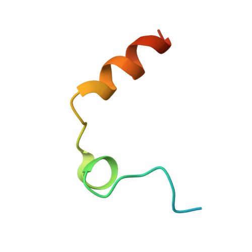 |
UniProt & NIH Common Fund Data Resources | |||||
Find proteins for P00734 (Homo sapiens) Explore P00734 Go to UniProtKB: P00734 | |||||
PHAROS: P00734 GTEx: ENSG00000180210 | |||||
Entity Groups | |||||
| Sequence Clusters | 30% Identity50% Identity70% Identity90% Identity95% Identity100% Identity | ||||
| UniProt Group | P00734 | ||||
Sequence AnnotationsExpand | |||||
| |||||
Find similar proteins by:
(by identity cutoff) | 3D Structure
Entity ID: 2 | |||||
|---|---|---|---|---|---|
| Molecule | Chains | Sequence Length | Organism | Details | Image |
| Prothrombin | B [auth H] | 259 | Homo sapiens | Mutation(s): 0 EC: 3.4.21.5 | 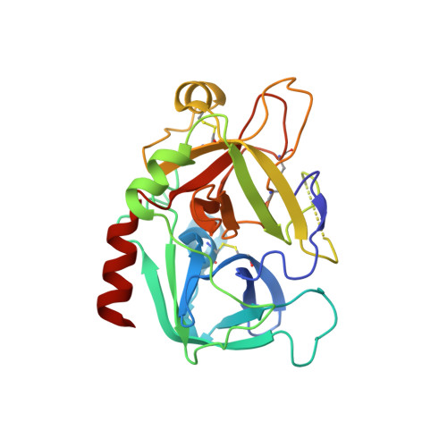 |
UniProt & NIH Common Fund Data Resources | |||||
Find proteins for P00734 (Homo sapiens) Explore P00734 Go to UniProtKB: P00734 | |||||
PHAROS: P00734 GTEx: ENSG00000180210 | |||||
Entity Groups | |||||
| Sequence Clusters | 30% Identity50% Identity70% Identity90% Identity95% Identity100% Identity | ||||
| UniProt Group | P00734 | ||||
Sequence AnnotationsExpand | |||||
| |||||
Find similar nucleic acids by: Sequence | 3D Structure
Entity ID: 3 | |||||
|---|---|---|---|---|---|
| Molecule | Chains | Length | Organism | Image | |
| Thrombin binding aptamer | C [auth D] | 15 | synthetic construct | 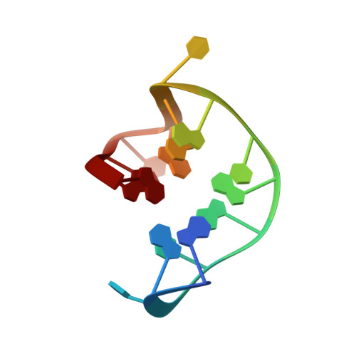 | |
Sequence AnnotationsExpand | |||||
| |||||
Small Molecules
| Ligands 6 Unique | |||||
|---|---|---|---|---|---|
| ID | Chains | Name / Formula / InChI Key | 2D Diagram | 3D Interactions | |
| 0G6 Query on 0G6 | E [auth H] | D-phenylalanyl-N-[(2S,3S)-6-{[amino(iminio)methyl]amino}-1-chloro-2-hydroxyhexan-3-yl]-L-prolinamide C21 H34 Cl N6 O3 DVFLYEYCMMLBTQ-VSZNYVQBSA-O |  | ||
| NAG Query on NAG | F [auth H] | 2-acetamido-2-deoxy-beta-D-glucopyranose C8 H15 N O6 OVRNDRQMDRJTHS-FMDGEEDCSA-N |  | ||
| ZN Query on ZN | D [auth L], H | ZINC ION Zn PTFCDOFLOPIGGS-UHFFFAOYSA-N |  | ||
| K Query on K | K [auth D] | POTASSIUM ION K NPYPAHLBTDXSSS-UHFFFAOYSA-N |  | ||
| CL Query on CL | I [auth H], J [auth H] | CHLORIDE ION Cl VEXZGXHMUGYJMC-UHFFFAOYSA-M |  | ||
| NA Query on NA | G [auth H] | SODIUM ION Na FKNQFGJONOIPTF-UHFFFAOYSA-N |  | ||
Biologically Interesting Molecules (External Reference) 1 Unique
Entity ID: 5 | |||||
|---|---|---|---|---|---|
| ID | Chains | Name | Type/Class | 2D Diagram | 3D Interactions |
| PRD_000020 (0G6) Query on PRD_000020 | E [auth H] | D-Phe-Pro-Arg-CH2Cl | Peptide-like / Inhibitor | | |
Experimental Data & Validation
Experimental Data
- Method: X-RAY DIFFRACTION
- Resolution: 2.05 Å
- R-Value Free: 0.226
- R-Value Work: 0.174
- R-Value Observed: 0.195
- Space Group: P 1
Unit Cell:
| Length ( Å ) | Angle ( ˚ ) |
|---|---|
| a = 43.369 | α = 68.97 |
| b = 45.299 | β = 86.11 |
| c = 51.519 | γ = 69.66 |
| Software Name | Purpose |
|---|---|
| CrystalClear | data collection |
| PHASES | phasing |
| CNS | refinement |
| HKL-2000 | data reduction |
| HKL-2000 | data scaling |
Entry History
Deposition Data
- Released Date: 2012-07-18 Deposition Author(s): Russo Krauss, I., Merlino, A., Mazzarella, L., Sica, F.
Revision History (Full details and data files)
- Version 1.0: 2012-07-18
Type: Initial release - Version 1.1: 2012-09-26
Changes: Database references - Version 1.2: 2013-02-27
Changes: Other - Version 1.3: 2020-07-29
Type: Remediation
Reason: Carbohydrate remediation
Changes: Data collection, Derived calculations, Structure summary - Version 1.4: 2023-09-13
Changes: Data collection, Database references, Refinement description, Structure summary
