4DCX

X-ray structure of NikA in complex with Fe(1R,2R)-N,N'-Bis(2-pyridylmethyl)-N,N'-dicarboxymethyl-1,2-cyclohexanediamine


Experimental Data Snapshot

  • Method: X-RAY DIFFRACTION
  • Resolution: 2.00 Å
  • R-Value Free: 0.203 
  • R-Value Work: 0.155 
  • R-Value Observed: 0.157 

wwPDB Validation   3D Report Full Report


Ligand Structure Quality Assessment 


This is version 1.2 of the entry. See complete history


Literature

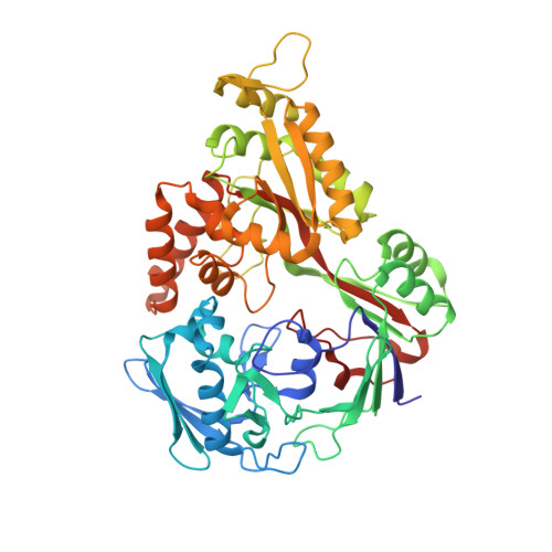
The structure of the periplasmic nickel-binding protein NikA provides insights for artificial metalloenzyme design.

Cherrier, M.V.Girgenti, E.Amara, P.Iannello, M.Marchi-Delapierre, C.Fontecilla-Camps, J.C.Menage, S.Cavazza, C.

(2012) J Biol Inorg Chem 17: 817-829

  • DOI: https://doi.org/10.1007/s00775-012-0899-7
  • Primary Citation of Related Structures:  
    4DCX, 4DCY

  • PubMed Abstract: 

    Understanding the interaction of a protein with a relevant ligand is crucial for the design of an artificial metalloenzyme. Our own interest is focused on the synthesis of artificial monooxygenases. In an initial effort, we have used the periplasmic nickel-binding protein NikA from Escherichia coli and iron complexes in which N(2)Py(2) ligands (where Py is pyridine) have been varied in terms of charge, aromaticity, and size. Six "NikA/iron complex" hybrids have been characterized by X-ray crystallography, and their interactions and solution properties have been studied. The hybrids are stable as indicated by their K (d) values, which are all in the micromolar range. The X-ray structures show that the ligands interact with NikA through salt bridges with arginine residues and π-stacking with a tryptophan residue. We have further characterized these interactions using quantum mechanical calculations and determined that weak CH/π hydrogen bonds finely modulate the stability differences between hybrids. We emphasize the important role of the tryptophan residues. Thus, our study aims at the complete characterization of the factors that condition the interaction of an artificial ligand and a protein and their implications for catalysis. Besides its potential usefulness in the synthesis of artificial monooxygenases, our approach should be generally applicable in the field of artificial metalloenzymes.


  • Organizational Affiliation

    Institut de Biologie Structurale Jean-Pierre Ebel, Groupe Métalloproteines, UMR 5075, CEA, CNRS, Université Joseph Fourier Grenoble 1, 41 rue Horowitz, 38027, Grenoble Cedex 1, France.


Macromolecules
Find similar proteins by:  (by identity cutoff)  |  3D Structure
Entity ID: 1
MoleculeChains Sequence LengthOrganismDetailsImage
Nickel-binding periplasmic protein
A, B
502Escherichia coli K-12Mutation(s): 0 
Gene Names: b3476JW3441nikA
UniProt
Find proteins for P33590 (Escherichia coli (strain K12))
Explore P33590 
Go to UniProtKB:  P33590
Entity Groups  
Sequence Clusters30% Identity50% Identity70% Identity90% Identity95% Identity100% Identity
UniProt GroupP33590
Sequence Annotations
Expand
  • Reference Sequence
Small Molecules
Ligands 4 Unique
IDChains Name / Formula / InChI Key2D Diagram3D Interactions
L2D
Query on L2D

Download Ideal Coordinates CCD File 
FA [auth A],
ZA [auth B]
{2,2'-[(1R,2R)-cyclohexane-1,2-diylbis{[(pyridin-2-yl-kappaN)methyl]imino-kappaN}]diacetato-kappaO(2-)}iron
C22 H26 Fe N4 O4
RHFVCFXUQWXGET-GZJHNZOKSA-L
SO4
Query on SO4

Download Ideal Coordinates CCD File 
C [auth A],
D [auth A],
GA [auth B]
SULFATE ION
O4 S
QAOWNCQODCNURD-UHFFFAOYSA-L
GOL
Query on GOL

Download Ideal Coordinates CCD File 
AA [auth A]
BA [auth A]
CA [auth A]
DA [auth A]
EA [auth A]
AA [auth A],
BA [auth A],
CA [auth A],
DA [auth A],
EA [auth A],
RA [auth B],
SA [auth B],
TA [auth B],
UA [auth B],
V [auth A],
VA [auth B],
W [auth A],
WA [auth B],
X [auth A],
XA [auth B],
Y [auth A],
YA [auth B],
Z [auth A]
GLYCEROL
C3 H8 O3
PEDCQBHIVMGVHV-UHFFFAOYSA-N
ACT
Query on ACT

Download Ideal Coordinates CCD File 
E [auth A]
F [auth A]
G [auth A]
H [auth A]
HA [auth B]
E [auth A],
F [auth A],
G [auth A],
H [auth A],
HA [auth B],
I [auth A],
IA [auth B],
J [auth A],
JA [auth B],
K [auth A],
KA [auth B],
L [auth A],
LA [auth B],
M [auth A],
MA [auth B],
N [auth A],
NA [auth B],
O [auth A],
OA [auth B],
P [auth A],
PA [auth B],
Q [auth A],
QA [auth B],
R [auth A],
S [auth A],
T [auth A],
U [auth A]
ACETATE ION
C2 H3 O2
QTBSBXVTEAMEQO-UHFFFAOYSA-M
Binding Affinity Annotations 
IDSourceBinding Affinity
L2D PDBBind:  4DCX Kd: 3.14e+4 (nM) from 1 assay(s)
Experimental Data & Validation

Experimental Data

  • Method: X-RAY DIFFRACTION
  • Resolution: 2.00 Å
  • R-Value Free: 0.203 
  • R-Value Work: 0.155 
  • R-Value Observed: 0.157 
  • Space Group: P 21 21 21
Unit Cell:
Length ( Å )Angle ( ˚ )
a = 86.35α = 90
b = 95.64β = 90
c = 125.19γ = 90
Software Package:
Software NamePurpose
PHASERphasing
PHENIXrefinement
XDSdata reduction
XSCALEdata scaling

Structure Validation

View Full Validation Report


Ligand Structure Quality Assessment 


Entry History 

Deposition Data

Revision History  (Full details and data files)

  • Version 1.0: 2012-05-09
    Type: Initial release
  • Version 1.1: 2012-06-13
    Changes: Database references
  • Version 1.2: 2023-09-13
    Changes: Data collection, Database references, Derived calculations, Refinement description