4CID

Structural insights into the N-terminus of the EHD2 ATPase


Experimental Data Snapshot

  • Method: X-RAY DIFFRACTION
  • Resolution: 3.00 Å
  • R-Value Free: 0.279 
  • R-Value Work: 0.230 
  • R-Value Observed: 0.233 

wwPDB Validation   3D Report Full Report


Ligand Structure Quality Assessment 


This is version 1.2 of the entry. See complete history


Literature

Structural Insights Into Membrane Interaction and Caveolar Targeting of Dynamin-Like Ehd2.

Shah, C.Hegde, B.G.Mor, B.Behrmann, E.Mielke, T.Moenke, G.Spahn, C.M.Lundmark, R.Daumke, O.Langen, R.

(2014) Structure 22: 409

  • DOI: https://doi.org/10.1016/j.str.2013.12.015
  • Primary Citation of Related Structures:  
    4CID

  • PubMed Abstract: 

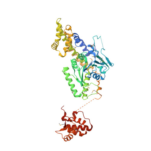
    The dynamin-related Eps15-homology domain-containing protein 2 (EHD2) is a membrane-remodeling ATPase that regulates the dynamics of caveolae. Here, we established an electron paramagnetic resonance (EPR) approach to characterize structural features of membrane-bound EHD2. We show that residues at the tip of the helical domain can insert into the membrane and may create membrane curvature by a wedging mechanism. Using EPR and X-ray crystallography, we found that the N terminus is folded into a hydrophobic pocket of the GTPase domain in solution and can be released into the membrane. Cryoelectron microscopy demonstrated that the N terminus is not essential for oligomerization of EHD2 into a membrane-anchored scaffold. Instead, we found a function of the N terminus in regulating targeting and stable association of EHD2 to caveolae. Our data uncover an unexpected, membrane-induced regulatory switch in EHD2 and demonstrate the versatility of EPR to study structure and function of dynamin superfamily proteins.


  • Organizational Affiliation

    Max-Delbrück-Center for Molecular Medicine, Crystallography, Robert-Rössle-Straße 10, 13092 Berlin, Germany.


Macromolecules
Find similar proteins by:  (by identity cutoff)  |  3D Structure
Entity ID: 1
MoleculeChains Sequence LengthOrganismDetailsImage
EH DOMAIN-CONTAINING PROTEIN 2550Mus musculusMutation(s): 2 
EC: 3.6.5.5
UniProt
Find proteins for Q8BH64 (Mus musculus)
Explore Q8BH64 
Go to UniProtKB:  Q8BH64
Entity Groups  
Sequence Clusters30% Identity50% Identity70% Identity90% Identity95% Identity100% Identity
UniProt GroupQ8BH64
Sequence Annotations
Expand
  • Reference Sequence
Small Molecules
Modified Residues  1 Unique
IDChains TypeFormula2D DiagramParent
MSE
Query on MSE
A
L-PEPTIDE LINKINGC5 H11 N O2 SeMET
Experimental Data & Validation

Experimental Data

  • Method: X-RAY DIFFRACTION
  • Resolution: 3.00 Å
  • R-Value Free: 0.279 
  • R-Value Work: 0.230 
  • R-Value Observed: 0.233 
  • Space Group: C 1 2 1
Unit Cell:
Length ( Å )Angle ( ˚ )
a = 99.851α = 90
b = 134.653β = 106.05
c = 56.068γ = 90
Software Package:
Software NamePurpose
XDSdata reduction
XDSdata scaling
SHARPphasing
REFMACphasing
REFMACrefinement

Structure Validation

View Full Validation Report


Ligand Structure Quality Assessment 


Entry History 

Deposition Data

Revision History  (Full details and data files)

  • Version 1.0: 2014-02-19
    Type: Initial release
  • Version 1.1: 2014-03-19
    Changes: Database references
  • Version 1.2: 2023-12-20
    Changes: Data collection, Database references, Derived calculations, Other, Refinement description