4BPT

Structural and thermodynamic insight into phenylalanine hydroxylase from the human pathogen Legionella pneumophila


Experimental Data Snapshot

  • Method: X-RAY DIFFRACTION
  • Resolution: 2.50 Å
  • R-Value Free: 0.302 
  • R-Value Work: 0.264 
  • R-Value Observed: 0.265 

wwPDB Validation   3D Report Full Report


This is version 1.3 of the entry. See complete history


Literature

Structural and Thermodynamic Insight Into Phenylalanine Hydroxylase from the Human Pathogen Legionella Pneumophila.

Leiros, H.S.Flydal, M.I.Martinez, A.

(2013) FEBS Open Bio 3: 370

  • DOI: https://doi.org/10.1016/j.fob.2013.08.006
  • Primary Citation of Related Structures:  
    4BPT

  • PubMed Abstract: 

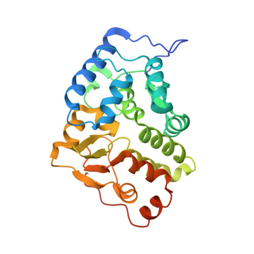
    Phenylalanine hydroxylase from Legionella pneumophila (lpPAH) has a major functional role in the synthesis of the pigment pyomelanin, which is a potential virulence factor. We present here the crystal structure of lpPAH, which is a dimeric enzyme that shows high thermostability, with a midpoint denaturation temperature of 79 °C, and low substrate affinity. The structure revealed a dimerization motif that includes ionic interactions and a hydrophobic core, composed of both β-structure and a C-terminal region, with the specific residues (P255, P256, Y257 and F258) interacting with the same residues from the adjacent subunit within the dimer. This unique dimerization interface, together with a number of aromatic clusters, appears to contribute to the high thermal stability of lpPAH. The crystal structure also explains the increased aggregation of the enzyme in the presence of salt. Moreover, the low affinity for substrate l-Phe could be explained from three consecutive glycine residues (G181, 182, 183) located at the substrate-binding site. This is the first structure of a dimeric bacterial PAH and provides a framework for interpreting the molecular and kinetic properties of lpPAH and for further investigating the regulation of the enzyme.


  • Organizational Affiliation

    The Norwegian Structural Biology Centre (NorStruct), Department of Chemistry, University of Tromsø, N-9037 Tromsø, Norway.


Macromolecules
Find similar proteins by:  (by identity cutoff)  |  3D Structure
Entity ID: 1
MoleculeChains Sequence LengthOrganismDetailsImage
PHENYLALANINE-4-HYDROXYLASE (PAH) (PHE-4-MONOOXYGENASE)
A, B, C, D
272Legionella pneumophilaMutation(s): 0 
EC: 1.14.16.1
UniProt
Find proteins for Q5ZS72 (Legionella pneumophila subsp. pneumophila (strain Philadelphia 1 / ATCC 33152 / DSM 7513))
Explore Q5ZS72 
Go to UniProtKB:  Q5ZS72
Entity Groups  
Sequence Clusters30% Identity50% Identity70% Identity90% Identity95% Identity100% Identity
UniProt GroupQ5ZS72
Sequence Annotations
Expand
  • Reference Sequence
Experimental Data & Validation

Experimental Data

  • Method: X-RAY DIFFRACTION
  • Resolution: 2.50 Å
  • R-Value Free: 0.302 
  • R-Value Work: 0.264 
  • R-Value Observed: 0.265 
  • Space Group: P 1 21 1
Unit Cell:
Length ( Å )Angle ( ˚ )
a = 90.32α = 90
b = 60.12β = 94.07
c = 124.04γ = 90
Software Package:
Software NamePurpose
REFMACrefinement
XDSdata reduction
XDSdata scaling
PHASERphasing

Structure Validation

View Full Validation Report


Entry History 

Deposition Data

Revision History  (Full details and data files)

  • Version 1.0: 2013-08-21
    Type: Initial release
  • Version 1.1: 2013-08-28
    Changes: Atomic model
  • Version 1.2: 2014-01-29
    Changes: Database references
  • Version 1.3: 2023-12-20
    Changes: Data collection, Database references, Derived calculations, Other, Refinement description