3C15

Complex of GS-Alpha with the Catalytic Domains of Mammalian Adenylyl Cyclase: Complex with Pyrophosphate and Mg


Experimental Data Snapshot

  • Method: X-RAY DIFFRACTION
  • Resolution: 2.78 Å
  • R-Value Free: 0.291 
  • R-Value Work: 0.237 
  • R-Value Observed: 0.240 

wwPDB Validation   3D Report Full Report


Ligand Structure Quality Assessment 


This is version 1.3 of the entry. See complete history


Literature

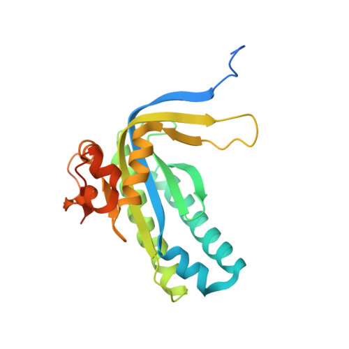
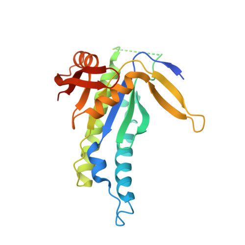
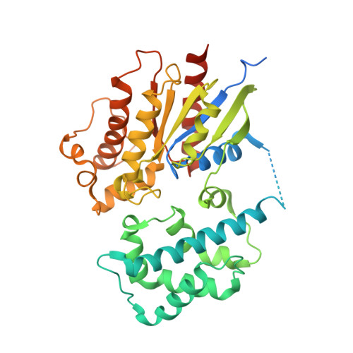
Structural basis for inhibition of mammalian adenylyl cyclase by calcium.

Mou, T.C.Masada, N.Cooper, D.M.Sprang, S.R.

(2009) Biochemistry 48: 3387-3397

  • DOI: https://doi.org/10.1021/bi802122k
  • Primary Citation of Related Structures:  
    3C14, 3C15, 3C16, 3MAA

  • PubMed Abstract: 

    Type V and VI mammalian adenylyl cyclases (AC5, AC6) are inhibited by Ca(2+) at both sub- and supramicromolar concentration. This inhibition may provide feedback in situations where cAMP promotes opening of Ca(2+) channels, allowing fine control of cardiac contraction and rhythmicity in cardiac tissue where AC5 and AC6 predominate. Ca(2+) inhibits the soluble AC core composed of the C1 domain of AC5 (VC1) and the C2 domain of AC2 (IIC2). As observed for holo-AC5, inhibition is biphasic, showing "high-affinity" (K(i) = approximately 0.4 microM) and "low-affinity" (K(i) = approximately 100 microM) modes of inhibition. At micromolar concentration, Ca(2+) inhibition is nonexclusive with respect to pyrophosphate (PP(i)), a noncompetitive inhibitor with respect to ATP, but at >100 microM Ca(2+), inhibition appears to be exclusive with respect to PP(i). The 3.0 A resolution structure of Galphas.GTPgammaS/forskolin-activated VC1:IIC2 crystals soaked in the presence of ATPalphaS and 8 microM free Ca(2+) contains a single, loosely coordinated metal ion. ATP soaked into VC1:IIC2 crystals in the presence of 1.5 mM Ca(2+) is not cyclized, and two calcium ions are observed in the 2.9 A resolution structure of the complex. In both of the latter complexes VC1:IIC2 adopts the "open", catalytically inactive conformation characteristic of the apoenzyme, in contrast to the "closed", active conformation seen in the presence of ATP analogues and Mg(2+) or Mn(2+). Structures of the pyrophosphate (PP(i)) complex with 10 mM Mg(2+) (2.8 A) or 2 mM Ca(2+) (2.7 A) also adopt the open conformation, indicating that the closed to open transition occurs after cAMP release. In the latter complexes, Ca(2+) and Mg(2+) bind only to the high-affinity "B" metal site associated with substrate/product stabilization. Ca(2+) thus stabilizes the inactive conformation in both ATP- and PP(i)-bound states.


  • Organizational Affiliation

    Center for Biomolecular Structure and Dynamics and Division of Biological Sciences, The University of Montana, Missoula, Montana 59812, USA.


Macromolecules
Find similar proteins by:  (by identity cutoff)  |  3D Structure
Entity ID: 1
MoleculeChains Sequence LengthOrganismDetailsImage
Adenylate cyclase type 5225Canis lupus familiarisMutation(s): 1 
Gene Names: ADCY5
EC: 4.6.1.1
UniProt
Find proteins for P30803 (Canis lupus familiaris)
Explore P30803 
Go to UniProtKB:  P30803
Entity Groups  
Sequence Clusters30% Identity50% Identity70% Identity90% Identity95% Identity100% Identity
UniProt GroupP30803
Sequence Annotations
Expand
  • Reference Sequence
Find similar proteins by:  (by identity cutoff)  |  3D Structure
Entity ID: 2
MoleculeChains Sequence LengthOrganismDetailsImage
Adenylate cyclase type 2212Rattus norvegicusMutation(s): 0 
Gene Names: Adcy2
EC: 4.6.1.1
UniProt
Find proteins for P26769 (Rattus norvegicus)
Explore P26769 
Go to UniProtKB:  P26769
Entity Groups  
Sequence Clusters30% Identity50% Identity70% Identity90% Identity95% Identity100% Identity
UniProt GroupP26769
Sequence Annotations
Expand
  • Reference Sequence
Find similar proteins by:  (by identity cutoff)  |  3D Structure
Entity ID: 3
MoleculeChains Sequence LengthOrganismDetailsImage
Guanine nucleotide-binding protein G(s) subunit alpha isoforms short402Bos taurusMutation(s): 0 
Gene Names: GNASGNAS1
UniProt
Find proteins for P04896 (Bos taurus)
Explore P04896 
Go to UniProtKB:  P04896
Entity Groups  
Sequence Clusters30% Identity50% Identity70% Identity90% Identity95% Identity100% Identity
UniProt GroupP04896
Sequence Annotations
Expand
  • Reference Sequence
Small Molecules
Ligands 5 Unique
IDChains Name / Formula / InChI Key2D Diagram3D Interactions
GSP
Query on GSP

Download Ideal Coordinates CCD File 
H [auth C]5'-GUANOSINE-DIPHOSPHATE-MONOTHIOPHOSPHATE
C10 H16 N5 O13 P3 S
XOFLBQFBSOEHOG-UUOKFMHZSA-N
FOK
Query on FOK

Download Ideal Coordinates CCD File 
E [auth A]FORSKOLIN
C22 H34 O7
OHCQJHSOBUTRHG-KGGHGJDLSA-N
POP
Query on POP

Download Ideal Coordinates CCD File 
F [auth A]PYROPHOSPHATE 2-
H2 O7 P2
XPPKVPWEQAFLFU-UHFFFAOYSA-L
CL
Query on CL

Download Ideal Coordinates CCD File 
G [auth C]CHLORIDE ION
Cl
VEXZGXHMUGYJMC-UHFFFAOYSA-M
MG
Query on MG

Download Ideal Coordinates CCD File 
D [auth A]MAGNESIUM ION
Mg
JLVVSXFLKOJNIY-UHFFFAOYSA-N
Experimental Data & Validation

Experimental Data

  • Method: X-RAY DIFFRACTION
  • Resolution: 2.78 Å
  • R-Value Free: 0.291 
  • R-Value Work: 0.237 
  • R-Value Observed: 0.240 
  • Space Group: P 21 21 2
Unit Cell:
Length ( Å )Angle ( ˚ )
a = 117.51α = 90
b = 133.08β = 90
c = 70.108γ = 90
Software Package:
Software NamePurpose
REFMACrefinement
HKL-2000data collection
HKL-2000data reduction
HKL-2000data scaling
PHASESphasing

Structure Validation

View Full Validation Report


Ligand Structure Quality Assessment 


Entry History 

Deposition Data

Revision History  (Full details and data files)

  • Version 1.0: 2009-02-03
    Type: Initial release
  • Version 1.1: 2011-07-13
    Changes: Version format compliance
  • Version 1.2: 2021-10-20
    Changes: Database references, Derived calculations
  • Version 1.3: 2023-08-30
    Changes: Data collection, Refinement description