3V3Z

I(L177)H mutant structure of photosynthetic reaction center from Rhodobacter sphaeroides


Experimental Data Snapshot

  • Method: X-RAY DIFFRACTION
  • Resolution: 2.90 Å
  • R-Value Free: 0.251 
  • R-Value Work: 0.204 
  • R-Value Observed: 0.206 

wwPDB Validation   3D Report Full Report


Ligand Structure Quality Assessment 


This is version 1.2 of the entry. See complete history


Literature

The site-directed mutation I(L177)H in Rhodobacter sphaeroides reaction center affects coordination of P(A) and B(B) bacteriochlorophylls.

Vasilieva, L.G.Fufina, T.Y.Gabdulkhakov, A.G.Leonova, M.M.Khatypov, R.A.Shuvalov, V.A.

(2012) Biochim Biophys Acta 1817: 1407-1417

  • DOI: https://doi.org/10.1016/j.bbabio.2012.02.008
  • Primary Citation of Related Structures:  
    3V3Y, 3V3Z

  • PubMed Abstract: 

    To explore the influence of the I(L177)H single mutation on the properties of the nearest bacteriochlorophylls (BChls), three reaction centers (RCs) bearing double mutations were constructed in the photosynthetic purple bacterium Rhodobacter sphaeroides, and their properties and pigment content were compared with those of the correspondent single mutant RCs. Each pair of the mutations comprised the amino acid substitution I(L177)H and another mutation altering histidine ligand of BChl P(A) or BChl B(B). Contrary to expectations, the double mutation I(L177)H+H(L173)L does not bring about a heterodimer RC but causes a 46nm blue shift of the long-wavelength P absorbance band. The histidine L177 or a water molecule were suggested as putative ligands for P(A) in the RC I(L177)H+H(L173)L although this would imply a reorientation of the His backbone and additional rearrangements in the primary donor environment or even a repositioning of the BChl dimer. The crystal structure of the mutant I(L177)H reaction center determined to a resolution of 2.9Å shows changes at the interface region between the BChl P(A) and the monomeric BChl B(B). Spectral and pigment analysis provided evidence for β-coordination of the BChl B(B) in the double mutant RC I(L177)H+H(M182)L and for its hexacoordination in the mutant reaction center I(L177)H. Computer modeling suggests involvement of two water molecules in the β-coordination of the BChl B(B). Possible structural consequences of the L177 mutation affecting the coordination of the two BChls P(A) and B(B) are discussed. This article is part of a Special Issue entitled: Photosynthesis Research for Sustainability: from Natural to Artificial.


  • Organizational Affiliation

    Institute of Basic Biological Problems, Russian Academy of Sciences, Pushchino, Moscow, Russia.


Macromolecules
Find similar proteins by:  (by identity cutoff)  |  3D Structure
Entity ID: 1
MoleculeChains Sequence LengthOrganismDetailsImage
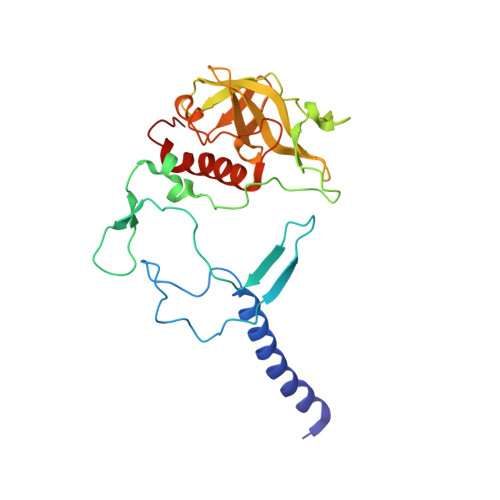
Reaction center protein H chainA [auth H]241Cereibacter sphaeroidesMutation(s): 0 
Gene Names: puhA
Membrane Entity: Yes 
UniProt
Find proteins for P0C0Y7 (Cereibacter sphaeroides)
Explore P0C0Y7 
Go to UniProtKB:  P0C0Y7
Entity Groups  
Sequence Clusters30% Identity50% Identity70% Identity90% Identity95% Identity100% Identity
UniProt GroupP0C0Y7
Sequence Annotations
Expand
  • Reference Sequence
Find similar proteins by:  (by identity cutoff)  |  3D Structure
Entity ID: 2
MoleculeChains Sequence LengthOrganismDetailsImage
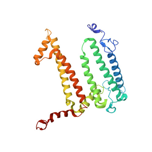
Reaction center protein L chainB [auth L]281Cereibacter sphaeroidesMutation(s): 1 
Gene Names: pufL
Membrane Entity: Yes 
UniProt
Find proteins for P0C0Y8 (Cereibacter sphaeroides)
Explore P0C0Y8 
Go to UniProtKB:  P0C0Y8
Entity Groups  
Sequence Clusters30% Identity50% Identity70% Identity90% Identity95% Identity100% Identity
UniProt GroupP0C0Y8
Sequence Annotations
Expand
  • Reference Sequence
Find similar proteins by:  (by identity cutoff)  |  3D Structure
Entity ID: 3
MoleculeChains Sequence LengthOrganismDetailsImage
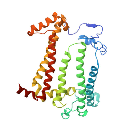
Reaction center protein M chainC [auth M]302Cereibacter sphaeroidesMutation(s): 0 
Gene Names: pufM
Membrane Entity: Yes 
UniProt
Find proteins for P0C0Y9 (Cereibacter sphaeroides)
Explore P0C0Y9 
Go to UniProtKB:  P0C0Y9
Entity Groups  
Sequence Clusters30% Identity50% Identity70% Identity90% Identity95% Identity100% Identity
UniProt GroupP0C0Y9
Sequence Annotations
Expand
  • Reference Sequence
Small Molecules
Ligands 11 Unique
IDChains Name / Formula / InChI Key2D Diagram3D Interactions
BCL
Query on BCL

Download Ideal Coordinates CCD File 
K [auth L],
L,
M [auth L],
R [auth M]
BACTERIOCHLOROPHYLL A
C55 H74 Mg N4 O6
DSJXIQQMORJERS-AGGZHOMASA-M
BPH
Query on BPH

Download Ideal Coordinates CCD File 
N [auth L],
S [auth M]
BACTERIOPHEOPHYTIN A
C55 H76 N4 O6
KWOZSBGNAHVCKG-SZQBJALDSA-N
U10
Query on U10

Download Ideal Coordinates CCD File 
O [auth L],
U [auth M]
UBIQUINONE-10
C59 H90 O4
ACTIUHUUMQJHFO-UPTCCGCDSA-N
SPN
Query on SPN

Download Ideal Coordinates CCD File 
V [auth M]SPEROIDENONE
C41 H70 O2
GWQAMGYOEYXWJF-YCDPMLDASA-N
LDA
Query on LDA

Download Ideal Coordinates CCD File 
D [auth H],
W [auth M],
X [auth M],
Y [auth M],
Z [auth M]
LAURYL DIMETHYLAMINE-N-OXIDE
C14 H31 N O
SYELZBGXAIXKHU-UHFFFAOYSA-N
HTO
Query on HTO

Download Ideal Coordinates CCD File 
G [auth H],
H,
P [auth L]
HEPTANE-1,2,3-TRIOL
C7 H16 O3
HXYCHJFUBNTKQR-RNFRBKRXSA-N
PO4
Query on PO4

Download Ideal Coordinates CCD File 
AA [auth M],
BA [auth M],
CA [auth M],
E [auth H],
F [auth H]
PHOSPHATE ION
O4 P
NBIIXXVUZAFLBC-UHFFFAOYSA-K
DIO
Query on DIO

Download Ideal Coordinates CCD File 
I [auth H],
Q [auth L]
1,4-DIETHYLENE DIOXIDE
C4 H8 O2
RYHBNJHYFVUHQT-UHFFFAOYSA-N
FE
Query on FE

Download Ideal Coordinates CCD File 
T [auth M]FE (III) ION
Fe
VTLYFUHAOXGGBS-UHFFFAOYSA-N
K
Query on K

Download Ideal Coordinates CCD File 
J [auth H]POTASSIUM ION
K
NPYPAHLBTDXSSS-UHFFFAOYSA-N
CL
Query on CL

Download Ideal Coordinates CCD File 
DA [auth M]CHLORIDE ION
Cl
VEXZGXHMUGYJMC-UHFFFAOYSA-M
Experimental Data & Validation

Experimental Data

  • Method: X-RAY DIFFRACTION
  • Resolution: 2.90 Å
  • R-Value Free: 0.251 
  • R-Value Work: 0.204 
  • R-Value Observed: 0.206 
  • Space Group: P 31 2 1
Unit Cell:
Length ( Å )Angle ( ˚ )
a = 140.13α = 90
b = 140.13β = 90
c = 186.03γ = 120
Software Package:
Software NamePurpose
XDSdata scaling
PHASERphasing
REFMACrefinement
XDSdata reduction

Structure Validation

View Full Validation Report


Ligand Structure Quality Assessment 


Entry History 

Deposition Data

Revision History  (Full details and data files)

  • Version 1.0: 2012-03-14
    Type: Initial release
  • Version 1.1: 2012-06-27
    Changes: Database references
  • Version 1.2: 2023-09-13
    Changes: Data collection, Database references, Derived calculations, Refinement description