3UIR

Crystal structure of the plasmin-textilinin-1 complex


Experimental Data Snapshot

  • Method: X-RAY DIFFRACTION
  • Resolution: 2.78 Å
  • R-Value Free: 0.258 
  • R-Value Work: 0.209 
  • R-Value Observed: 0.211 

wwPDB Validation   3D Report Full Report


This is version 1.1 of the entry. See complete history


Literature

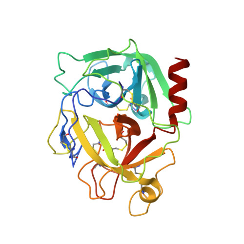
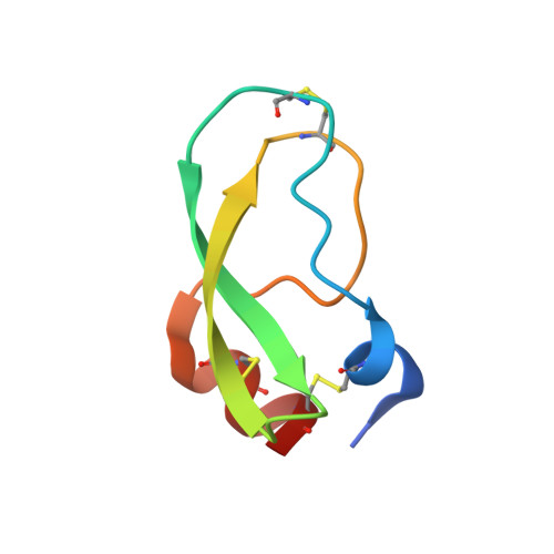
The structure of human microplasmin in complex with textilinin-1, an aprotinin-like inhibitor from the Australian brown snake.

Millers, E.K.Johnson, L.A.Birrell, G.W.Masci, P.P.Lavin, M.F.de Jersey, J.Guddat, L.W.

(2013) PLoS One 8: e54104-e54104

  • DOI: https://doi.org/10.1371/journal.pone.0054104
  • Primary Citation of Related Structures:  
    3UIR

  • PubMed Abstract: 

    Textilinin-1 is a Kunitz-type serine protease inhibitor from Australian brown snake venom. Its ability to potently and specifically inhibit human plasmin (K(i) = 0.44 nM) makes it a potential therapeutic drug as a systemic anti-bleeding agent. The crystal structures of the human microplasmin-textilinin-1 and the trypsin-textilinin-1 complexes have been determined to 2.78 Å and 1.64 Å resolution respectively, and show that textilinin-1 binds to trypsin in a canonical mode but to microplasmin in an atypical mode with the catalytic histidine of microplasmin rotated out of the active site. The space vacated by the histidine side-chain in this complex is partially occupied by a water molecule. In the structure of microplasminogen the χ(1) dihedral angle of the side-chain of the catalytic histidine is rotated by 67° from its "active" position in the catalytic triad, as exemplified by its location when microplasmin is bound to streptokinase. However, when textilinin-1 binds to microplasmin the χ(1) dihedral angle of this amino acid residue changes by -157° (i.e. in the opposite rotation direction compared to microplasminogen). The unusual mode of interaction between textilinin-1 and plasmin explains textilinin-1's selectivity for human plasmin over plasma kallikrein. This difference can be exploited in future drug design efforts.


  • Organizational Affiliation

    School of Chemistry and Molecular Biosciences, The University of Queensland, Brisbane, Queensland, Australia.


Macromolecules
Find similar proteins by:  (by identity cutoff)  |  3D Structure
Entity ID: 1
MoleculeChains Sequence LengthOrganismDetailsImage
Plasmin light chain B
A, B
247Homo sapiensMutation(s): 0 
Gene Names: PLG
EC: 3.4.21.7
UniProt & NIH Common Fund Data Resources
Find proteins for P00747 (Homo sapiens)
Explore P00747 
Go to UniProtKB:  P00747
PHAROS:  P00747
GTEx:  ENSG00000122194 
Entity Groups  
Sequence Clusters30% Identity50% Identity70% Identity90% Identity95% Identity100% Identity
UniProt GroupP00747
Sequence Annotations
Expand
  • Reference Sequence
Find similar proteins by:  (by identity cutoff)  |  3D Structure
Entity ID: 2
MoleculeChains Sequence LengthOrganismDetailsImage
Textilinin-1
C, D
59Pseudonaja textilis textilisMutation(s): 0 
UniProt
Find proteins for Q90WA1 (Pseudonaja textilis textilis)
Explore Q90WA1 
Go to UniProtKB:  Q90WA1
Entity Groups  
Sequence Clusters30% Identity50% Identity70% Identity90% Identity95% Identity100% Identity
UniProt GroupQ90WA1
Sequence Annotations
Expand
  • Reference Sequence
Experimental Data & Validation

Experimental Data

  • Method: X-RAY DIFFRACTION
  • Resolution: 2.78 Å
  • R-Value Free: 0.258 
  • R-Value Work: 0.209 
  • R-Value Observed: 0.211 
  • Space Group: P 1 21 1
Unit Cell:
Length ( Å )Angle ( ˚ )
a = 81.901α = 90
b = 48.019β = 102.08
c = 82.627γ = 90
Software Package:
Software NamePurpose
CrystalCleardata collection
EPMRphasing
PHENIXrefinement
CrystalCleardata reduction
CrystalCleardata scaling

Structure Validation

View Full Validation Report


Entry History 

Deposition Data

Revision History  (Full details and data files)

  • Version 1.0: 2012-12-26
    Type: Initial release
  • Version 1.1: 2013-07-17
    Changes: Database references