3U01

Crystal structure of onconase double mutant C30A/C75A at 1.12 A resolution


Experimental Data Snapshot

  • Method: X-RAY DIFFRACTION
  • Resolution: 1.12 Å
  • R-Value Free: 0.150 
  • R-Value Work: 0.136 
  • R-Value Observed: 0.137 

wwPDB Validation   3D Report Full Report


This is version 2.0 of the entry. See complete history


Literature

Investigating the effects of double mutation C30A/C75A on onconase structure: Studies at atomic resolution.

Kurpiewska, K.Torrent, G.Ribo, M.Loch, J.I.Vilanova, M.Lewinski, K.

(2014) Biopolymers 101: 454-460

  • DOI: https://doi.org/10.1002/bip.22403
  • Primary Citation of Related Structures:  
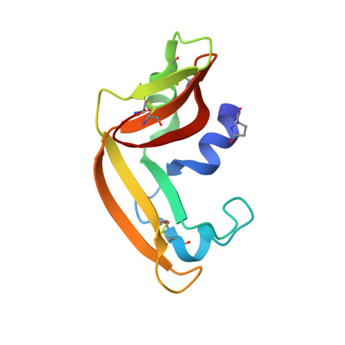
    3U01

  • PubMed Abstract: 

    The structure of onconase C30A/C75A double mutant has been determined at 1.12Å resolution. The structure has high structural homology to other onconase structures. The changes being results of mutation are relatively small, distributed asymmetrically around the two mutated positions, and they are observed not only in the mutation region but expanded to entire molecule. Different conformation of Lys31 side chain that influences the hydrogen bonding network around catalytic triad is probably responsible for lower catalytic efficiency of double mutant. The decrease in thermal stability observed for the onconase variant might be explained by a less dense packing as manifested by the increase of the molecular volume and the solvent accessible surface area.


  • Organizational Affiliation

    Faculty of Chemistry, Department of Crystal Physics and Crystal Chemistry, Protein Crystallography Group, Jagiellonian University, Ingardena 3, Kraków, 30060, Poland.


Macromolecules
Find similar proteins by:  (by identity cutoff)  |  3D Structure
Entity ID: 1
MoleculeChains Sequence LengthOrganismDetailsImage
Protein P-30104Lithobates pipiensMutation(s): 2 
EC: 3.1.27
UniProt
Find proteins for P22069 (Lithobates pipiens)
Explore P22069 
Go to UniProtKB:  P22069
Entity Groups  
Sequence Clusters30% Identity50% Identity70% Identity90% Identity95% Identity100% Identity
UniProt GroupP22069
Sequence Annotations
Expand
  • Reference Sequence
Experimental Data & Validation

Experimental Data

  • Method: X-RAY DIFFRACTION
  • Resolution: 1.12 Å
  • R-Value Free: 0.150 
  • R-Value Work: 0.136 
  • R-Value Observed: 0.137 
  • Space Group: P 21 21 21
Unit Cell:
Length ( Å )Angle ( ˚ )
a = 32.288α = 90
b = 38.664β = 90
c = 69.514γ = 90
Software Package:
Software NamePurpose
SCALAdata scaling
PHASERphasing
REFMACrefinement
PDB_EXTRACTdata extraction
CrysalisProdata collection
CrysalisProdata reduction

Structure Validation

View Full Validation Report


Entry History 

Deposition Data

Revision History  (Full details and data files)

  • Version 1.0: 2011-12-21
    Type: Initial release
  • Version 1.1: 2014-10-15
    Changes: Database references
  • Version 1.2: 2017-11-08
    Changes: Refinement description
  • Version 2.0: 2023-09-13
    Changes: Data collection, Database references, Derived calculations, Polymer sequence, Refinement description