3TJE

Crystal structure of Fas receptor extracellular domain in complex with Fab E09


Experimental Data Snapshot

  • Method: X-RAY DIFFRACTION
  • Resolution: 1.93 Å
  • R-Value Free: 0.232 
  • R-Value Work: 0.204 
  • R-Value Observed: 0.206 

wwPDB Validation   3D Report Full Report


This is version 1.3 of the entry. See complete history


Literature

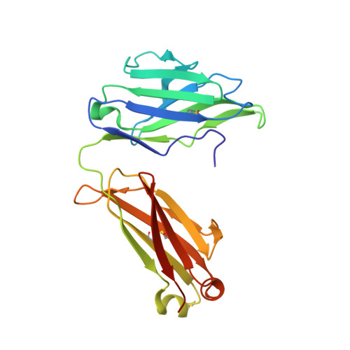
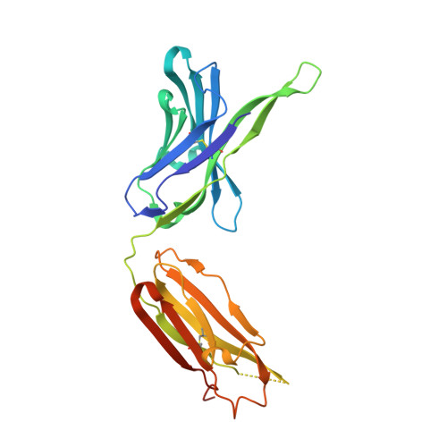
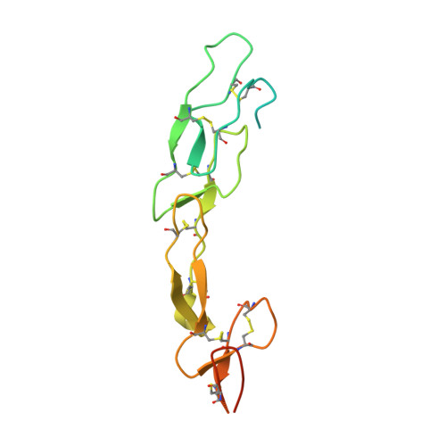
A series of Fas receptor agonist antibodies that demonstrate an inverse correlation between affinity and potency.

Chodorge, M.Zuger, S.Stirnimann, C.Briand, C.Jermutus, L.Grutter, M.G.Minter, R.R.

(2012) Cell Death Differ 19: 1187-1195

  • DOI: https://doi.org/10.1038/cdd.2011.208
  • Primary Citation of Related Structures:  
    3THM, 3TJE

  • PubMed Abstract: 

    Receptor agonism remains poorly understood at the molecular and mechanistic level. In this study, we identified a fully human anti-Fas antibody that could efficiently trigger apoptosis and therefore function as a potent agonist. Protein engineering and crystallography were used to mechanistically understand the agonistic activity of the antibody. The crystal structure of the complex was determined at 1.9 Å resolution and provided insights into epitope recognition and comparisons with the natural ligand FasL (Fas ligand). When we affinity-matured the agonist antibody, we observed that, surprisingly, the higher-affinity antibodies demonstrated a significant reduction, rather than an increase, in agonist activity at the Fas receptor. We propose and experimentally demonstrate a model to explain this non-intuitive impact of affinity on agonist antibody signalling and explore the implications for the discovery of therapeutic agonists in general.


  • Organizational Affiliation

    MedImmune Ltd., Granta Park, Cambridge, UK.


Macromolecules
Find similar proteins by:  (by identity cutoff)  |  3D Structure
Entity ID: 1
MoleculeChains Sequence LengthOrganismDetailsImage
Fab E09, light chainA [auth L]216Homo sapiensMutation(s): 0 
Entity Groups  
Sequence Clusters30% Identity50% Identity70% Identity90% Identity95% Identity100% Identity
Sequence Annotations
Expand
  • Reference Sequence
Find similar proteins by:  (by identity cutoff)  |  3D Structure
Entity ID: 2
MoleculeChains Sequence LengthOrganismDetailsImage
Fab E09, heavy chainB [auth H]245Homo sapiensMutation(s): 0 
Entity Groups  
Sequence Clusters30% Identity50% Identity70% Identity90% Identity95% Identity100% Identity
Sequence Annotations
Expand
  • Reference Sequence
Find similar proteins by:  (by identity cutoff)  |  3D Structure
Entity ID: 3
MoleculeChains Sequence LengthOrganismDetailsImage
Tumor necrosis factor receptor superfamily member 6C [auth F]156Homo sapiensMutation(s): 0 
Gene Names: APT1FASFAS1TNFRSF6
UniProt & NIH Common Fund Data Resources
Find proteins for P25445 (Homo sapiens)
Explore P25445 
Go to UniProtKB:  P25445
PHAROS:  P25445
GTEx:  ENSG00000026103 
Entity Groups  
Sequence Clusters30% Identity50% Identity70% Identity90% Identity95% Identity100% Identity
UniProt GroupP25445
Sequence Annotations
Expand
  • Reference Sequence
Experimental Data & Validation

Experimental Data

  • Method: X-RAY DIFFRACTION
  • Resolution: 1.93 Å
  • R-Value Free: 0.232 
  • R-Value Work: 0.204 
  • R-Value Observed: 0.206 
  • Space Group: C 2 2 21
Unit Cell:
Length ( Å )Angle ( ˚ )
a = 89.49α = 90
b = 166.4β = 90
c = 110.72γ = 90
Software Package:
Software NamePurpose
XSCALEdata scaling
PHASERphasing
PHENIXrefinement
PDB_EXTRACTdata extraction

Structure Validation

View Full Validation Report


Entry History 

Deposition Data

Revision History  (Full details and data files)

  • Version 1.0: 2012-05-09
    Type: Initial release
  • Version 1.1: 2012-06-27
    Changes: Database references
  • Version 1.2: 2017-11-08
    Changes: Refinement description
  • Version 1.3: 2023-09-13
    Changes: Data collection, Database references, Derived calculations, Refinement description