3STB

A complex of two editosome proteins and two nanobodies


Experimental Data Snapshot

  • Method: X-RAY DIFFRACTION
  • Resolution: 2.50 Å
  • R-Value Free: 0.234 
  • R-Value Work: 0.199 
  • R-Value Observed: 0.201 

wwPDB Validation   3D Report Full Report


This is version 1.4 of the entry. See complete history


Literature

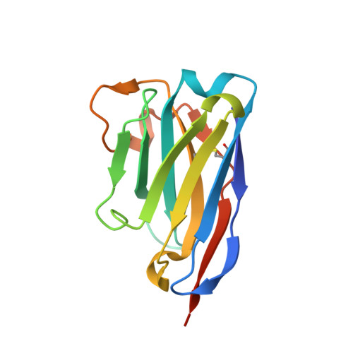
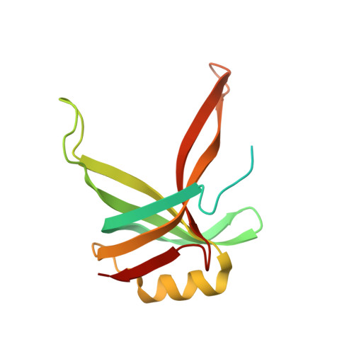
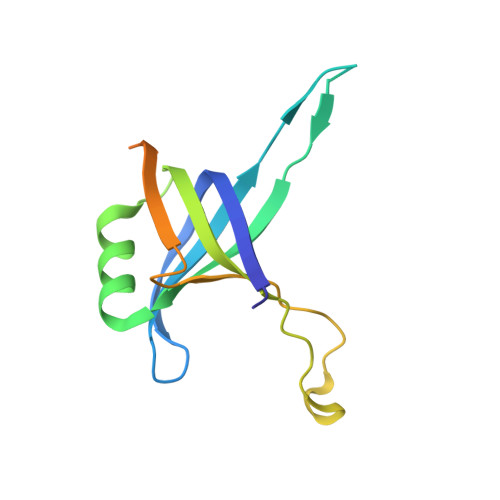
Crystal structure of a heterodimer of editosome interaction proteins in complex with two copies of a cross-reacting nanobody.

Park, Y.J.Pardon, E.Wu, M.Steyaert, J.Hol, W.G.

(2012) Nucleic Acids Res 40: 1828-1840

  • DOI: https://doi.org/10.1093/nar/gkr867
  • Primary Citation of Related Structures:  
    3STB

  • PubMed Abstract: 

    The parasite Trypanosoma brucei, the causative agent of sleeping sickness across sub-Saharan Africa, depends on a remarkable U-insertion/deletion RNA editing process in its mitochondrion. A approximately 20 S multi-protein complex, called the editosome, is an essential machinery for editing pre-mRNA molecules encoding the majority of mitochondrial proteins. Editosomes contain a common core of twelve proteins where six OB-fold interaction proteins, called A1-A6, play a crucial role. Here, we report the structure of two single-strand nucleic acid-binding OB-folds from interaction proteins A3 and A6 that surprisingly, form a heterodimer. Crystal growth required the assistance of an anti-A3 nanobody as a crystallization chaperone. Unexpectedly, this anti-A3 nanobody binds to both A3(OB) and A6, despite only ~40% amino acid sequence identity between the OB-folds of A3 and A6. The A3(OB)-A6 heterodimer buries 35% more surface area than the A6 homodimer. This is attributed mainly to the presence of a conserved Pro-rich loop in A3(OB). The implications of the A3(OB)-A6 heterodimer, and of a dimer of heterodimers observed in the crystals, for the architecture of the editosome are profound, resulting in a proposal of a 'five OB-fold center' in the core of the editosome.


  • Organizational Affiliation

    Department of Biochemistry, Biomolecular Structure Center, School of Medicine, University of Washington, PO Box 357742, Seattle WA 98195, USA.


Macromolecules
Find similar proteins by:  (by identity cutoff)  |  3D Structure
Entity ID: 1
MoleculeChains Sequence LengthOrganismDetailsImage
single domain antibody VHH
A, B
132Lama glamaMutation(s): 0 
Gene Names: single domain antibody VHH
Entity Groups  
Sequence Clusters30% Identity50% Identity70% Identity90% Identity95% Identity100% Identity
Sequence Annotations
Expand
  • Reference Sequence
Find similar proteins by:  (by identity cutoff)  |  3D Structure
Entity ID: 2
MoleculeChains Sequence LengthOrganismDetailsImage
RNA-editing complex protein MP42148Trypanosoma bruceiMutation(s): 0 
Gene Names: KREPA3Tb927.8.620
UniProt
Find proteins for Q95W13 (Trypanosoma brucei)
Explore Q95W13 
Go to UniProtKB:  Q95W13
Entity Groups  
Sequence Clusters30% Identity50% Identity70% Identity90% Identity95% Identity100% Identity
UniProt GroupQ95W13
Sequence Annotations
Expand
  • Reference Sequence
Find similar proteins by:  (by identity cutoff)  |  3D Structure
Entity ID: 3
MoleculeChains Sequence LengthOrganismDetailsImage
MP18 RNA editing complex protein145Trypanosoma bruceiMutation(s): 0 
Gene Names: KREPA6Tb10.70.2090
UniProt
Find proteins for Q38B90 (Trypanosoma brucei brucei (strain 927/4 GUTat10.1))
Explore Q38B90 
Go to UniProtKB:  Q38B90
Entity Groups  
Sequence Clusters30% Identity50% Identity70% Identity90% Identity95% Identity100% Identity
UniProt GroupQ38B90
Sequence Annotations
Expand
  • Reference Sequence
Experimental Data & Validation

Experimental Data

  • Method: X-RAY DIFFRACTION
  • Resolution: 2.50 Å
  • R-Value Free: 0.234 
  • R-Value Work: 0.199 
  • R-Value Observed: 0.201 
  • Space Group: P 43 21 2
Unit Cell:
Length ( Å )Angle ( ˚ )
a = 74.54α = 90
b = 74.54β = 90
c = 239.89γ = 90
Software Package:
Software NamePurpose
REFMACrefinement
PHASESphasing
PHENIXrefinement
HKL-2000data reduction
SCALAdata scaling
HKL-2000data collection

Structure Validation

View Full Validation Report


Entry History 

Deposition Data

Revision History  (Full details and data files)

  • Version 1.0: 2011-11-02
    Type: Initial release
  • Version 1.1: 2011-11-23
    Changes: Database references
  • Version 1.2: 2012-01-11
    Changes: Database references
  • Version 1.3: 2012-03-14
    Changes: Database references
  • Version 1.4: 2019-07-17
    Changes: Data collection, Refinement description