3RBF

Crystal structure of Human aromatic L-amino acid decarboxylase (AADC) in the apo form


Experimental Data Snapshot

  • Method: X-RAY DIFFRACTION
  • Resolution: 2.90 Å
  • R-Value Free: 0.267 
  • R-Value Work: 0.212 
  • R-Value Observed: 0.215 

wwPDB Validation   3D Report Full Report


Ligand Structure Quality Assessment 


This is version 1.3 of the entry. See complete history


Literature

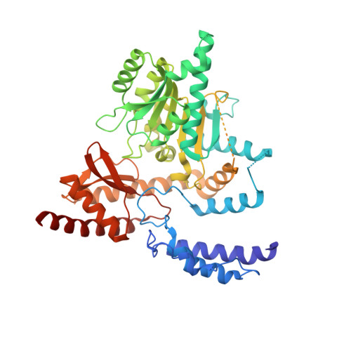
Open conformation of human DOPA decarboxylase reveals the mechanism of PLP addition to Group II decarboxylases.

Giardina, G.Montioli, R.Gianni, S.Cellini, B.Paiardini, A.Voltattorni, C.B.Cutruzzola, F.

(2011) Proc Natl Acad Sci U S A 108: 20514-20519

  • DOI: https://doi.org/10.1073/pnas.1111456108
  • Primary Citation of Related Structures:  
    3RBF, 3RBL, 3RCH

  • PubMed Abstract: 

    DOPA decarboxylase, the dimeric enzyme responsible for the synthesis of neurotransmitters dopamine and serotonin, is involved in severe neurological diseases such as Parkinson disease, schizophrenia, and depression. Binding of the pyridoxal-5'-phosphate (PLP) cofactor to the apoenzyme is thought to represent a central mechanism for the regulation of its activity. We solved the structure of the human apoenzyme and found it exists in an unexpected open conformation: compared to the pig kidney holoenzyme, the dimer subunits move 20 Å apart and the two active sites become solvent exposed. Moreover, by tuning the PLP concentration in the crystals, we obtained two more structures with different conformations of the active site. Analysis of three-dimensional data coupled to a kinetic study allows to identify the structural determinants of the open/close conformational change occurring upon PLP binding and thereby propose a model for the preferential degradation of the apoenzymes of Group II decarboxylases.


  • Organizational Affiliation

    Dipartimento di Scienze Biochimiche A. Rossi Fanelli, Sapienza Università di Roma, Piazzale Aldo Moro 5, 00185 Rome, Italy.


Macromolecules
Find similar proteins by:  (by identity cutoff)  |  3D Structure
Entity ID: 1
MoleculeChains Sequence LengthOrganismDetailsImage
Aromatic-L-amino-acid decarboxylase
A, B
480Homo sapiensMutation(s): 0 
Gene Names: AADCDDChCG_1811384tcag7.584
EC: 4.1.1.28
UniProt & NIH Common Fund Data Resources
Find proteins for P20711 (Homo sapiens)
Explore P20711 
Go to UniProtKB:  P20711
PHAROS:  P20711
GTEx:  ENSG00000132437 
Entity Groups  
Sequence Clusters30% Identity50% Identity70% Identity90% Identity95% Identity100% Identity
UniProt GroupP20711
Sequence Annotations
Expand
  • Reference Sequence
Experimental Data & Validation

Experimental Data

  • Method: X-RAY DIFFRACTION
  • Resolution: 2.90 Å
  • R-Value Free: 0.267 
  • R-Value Work: 0.212 
  • R-Value Observed: 0.215 
  • Space Group: P 41 21 2
Unit Cell:
Length ( Å )Angle ( ˚ )
a = 175.84α = 90
b = 175.84β = 90
c = 74.96γ = 90
Software Package:
Software NamePurpose
MOSFLMdata reduction
SCALEPACKdata scaling
MOLREPphasing
REFMACrefinement
PDB_EXTRACTdata extraction
MAR345data collection

Structure Validation

View Full Validation Report


Ligand Structure Quality Assessment 


Entry History 

Deposition Data

Revision History  (Full details and data files)

  • Version 1.0: 2011-10-19
    Type: Initial release
  • Version 1.1: 2012-01-04
    Changes: Database references
  • Version 1.2: 2017-11-08
    Changes: Refinement description
  • Version 1.3: 2023-09-13
    Changes: Data collection, Database references, Derived calculations, Refinement description