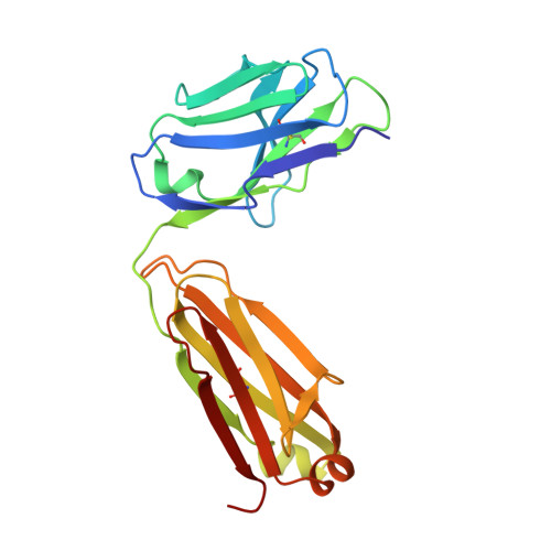
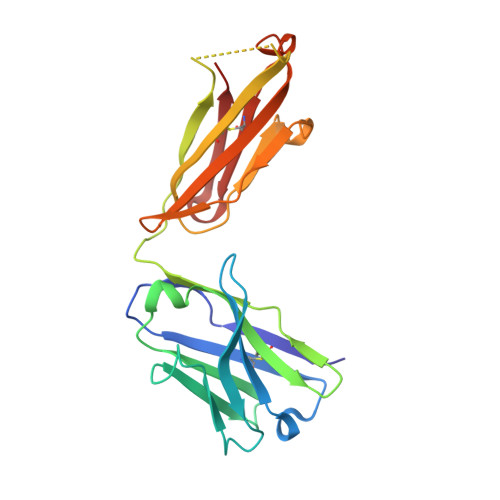
T cell receptors are structures capable of initiating signaling in the absence of large conformational rearrangements.
Fernandes, R.A., Shore, D.A., Vuong, M.T., Yu, C., Zhu, X., Pereira-Lopes, S., Brouwer, H., Fennelly, J.A., Jessup, C.M., Evans, E.J., Wilson, I.A., Davis, S.J.(2012) J Biol Chem 287: 13324-13335
- PubMed: 22262845
- DOI: https://doi.org/10.1074/jbc.M111.332783
- Primary Citation of Related Structures:
3R06, 3R08 - PubMed Abstract:
Native and non-native ligands of the T cell receptor (TCR), including antibodies, have been proposed to induce signaling in T cells via intra- or intersubunit conformational rearrangements within the extracellular regions of TCR complexes. We have investigated whether any signatures can be found for such postulated structural changes during TCR triggering induced by antibodies, using crystallographic and mutagenesis-based approaches. The crystal structure of murine CD3ε complexed with the mitogenic anti-CD3ε antibody 2C11 enabled the first direct structural comparisons of antibody-liganded and unliganded forms of CD3ε from a single species, which revealed that antibody binding does not induce any substantial rearrangements within CD3ε. Saturation mutagenesis of surface-exposed CD3ε residues, coupled with assays of antibody-induced signaling by the mutated complexes, suggests a new configuration for the complex within which CD3ε is highly exposed and reveals that no large new CD3ε interfaces are required to form during antibody-induced signaling. The TCR complex therefore appears to be a structure that is capable of initiating intracellular signaling in T cells without substantial structural rearrangements within or between the component subunits. Our findings raise the possibility that signaling by native ligands might also be initiated in the absence of large structural rearrangements in the receptor.
Organizational Affiliation:
Nuffield Department of Clinical Medicine and Medical Research Council Human Immunology Unit, The University of Oxford, John Radcliffe Hospital, Headington, Oxford OX3 9DU, United Kingdom.