3QQE

Crystal structure of HA2 R106H mutant of H2 hemagglutinin, re-neutralized form


Experimental Data Snapshot

  • Method: X-RAY DIFFRACTION
  • Resolution: 2.10 Å
  • R-Value Free: 0.235 
  • R-Value Work: 0.189 
  • R-Value Observed: 0.191 

wwPDB Validation   3D Report Full Report


Ligand Structure Quality Assessment 


This is version 2.0 of the entry. See complete history


Literature

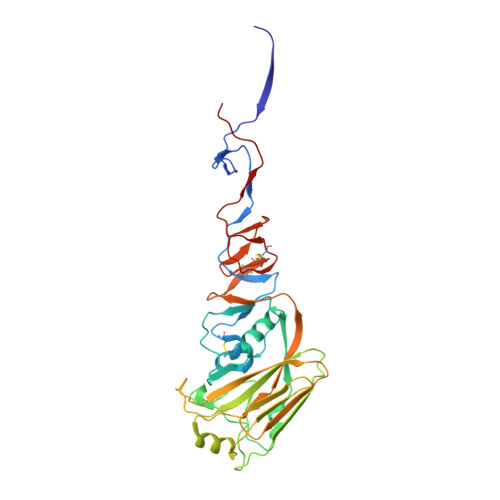
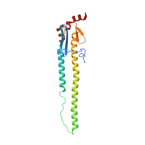
Structural Characterization of an Early Fusion Intermediate of Influenza Virus Hemagglutinin.

Xu, R.Wilson, I.A.

(2011) J Virol 85: 5172-5182

  • DOI: https://doi.org/10.1128/JVI.02430-10
  • Primary Citation of Related Structures:  
    3QQB, 3QQE, 3QQI, 3QQO

  • PubMed Abstract: 

    The hemagglutinin (HA) envelope protein of influenza virus mediates viral entry through membrane fusion in the acidic environment of the endosome. Crystal structures of HA in pre- and postfusion states have laid the foundation for proposals for a general fusion mechanism for viral envelope proteins. The large-scale conformational rearrangement of HA at low pH is triggered by a loop-to-helix transition of an interhelical loop (B loop) within the fusion domain and is often referred to as the "spring-loaded" mechanism. Although the receptor-binding HA1 subunit is believed to act as a "clamp" to keep the B loop in its metastable prefusion state at neutral pH, the "pH sensors" that are responsible for the clamp release and the ensuing structural transitions have remained elusive. Here we identify a mutation in the HA2 fusion domain from the influenza virus H2 subtype that stabilizes the HA trimer in a prefusion-like state at and below fusogenic pH. Crystal structures of this putative early intermediate state reveal reorganization of ionic interactions at the HA1-HA2 interface at acidic pH and deformation of the HA1 membrane-distal domain. Along with neutralization of glutamate residues on the B loop, these changes cause a rotation of the B loop and solvent exposure of conserved phenylalanines, which are key residues at the trimer interface of the postfusion structure. Thus, our study reveals the possible initial structural event that leads to release of the B loop from its prefusion conformation, which is aided by unexpected structural changes within the membrane-distal HA1 domain at low pH.


  • Organizational Affiliation

    Department of Molecular Biology, BCC-206, The Scripps Research Institute, 10550 North Torrey Pines Road, La Jolla, CA 92037, USA.


Macromolecules
Find similar proteins by:  (by identity cutoff)  |  3D Structure
Entity ID: 1
MoleculeChains Sequence LengthOrganismDetailsImage
Hemagglutinin327Influenza A virus (A/Japan/305/1957(H2N2))Mutation(s): 0 
Gene Names: HAhemagglutinin
UniProt
Find proteins for C7S226 (Influenza A virus (strain A/Japan/305/1957 H2N2))
Explore C7S226 
Go to UniProtKB:  C7S226
Entity Groups  
Sequence Clusters30% Identity50% Identity70% Identity90% Identity95% Identity100% Identity
UniProt GroupC7S226
Sequence Annotations
Expand
  • Reference Sequence
Find similar proteins by:  (by identity cutoff)  |  3D Structure
Entity ID: 2
MoleculeChains Sequence LengthOrganismDetailsImage
Hemagglutinin174Influenza A virus (A/Japan/305/1957(H2N2))Mutation(s): 1 
Gene Names: HAhemagglutinin
UniProt
Find proteins for C7S226 (Influenza A virus (strain A/Japan/305/1957 H2N2))
Explore C7S226 
Go to UniProtKB:  C7S226
Entity Groups  
Sequence Clusters30% Identity50% Identity70% Identity90% Identity95% Identity100% Identity
UniProt GroupC7S226
Sequence Annotations
Expand
  • Reference Sequence
Oligosaccharides

Help

Entity ID: 3
MoleculeChains Length2D Diagram Glycosylation3D Interactions
2-acetamido-2-deoxy-beta-D-glucopyranose-(1-4)-2-acetamido-2-deoxy-beta-D-glucopyranose
C
2N-Glycosylation
Glycosylation Resources
GlyTouCan:  G42666HT
GlyCosmos:  G42666HT
GlyGen:  G42666HT
Experimental Data & Validation

Experimental Data

  • Method: X-RAY DIFFRACTION
  • Resolution: 2.10 Å
  • R-Value Free: 0.235 
  • R-Value Work: 0.189 
  • R-Value Observed: 0.191 
  • Space Group: P 63
Unit Cell:
Length ( Å )Angle ( ˚ )
a = 70.339α = 90
b = 70.339β = 90
c = 237.093γ = 120
Software Package:
Software NamePurpose
DENZOdata reduction
SCALEPACKdata scaling
PHASERphasing
REFMACrefinement
PDB_EXTRACTdata extraction
HKL-2000data collection
HKL-2000data reduction
HKL-2000data scaling

Structure Validation

View Full Validation Report


Ligand Structure Quality Assessment 


Entry History 

Deposition Data

Revision History  (Full details and data files)

  • Version 1.0: 2011-03-09
    Type: Initial release
  • Version 1.1: 2011-07-13
    Changes: Version format compliance
  • Version 1.2: 2017-11-08
    Changes: Refinement description
  • Version 2.0: 2020-07-29
    Type: Remediation
    Reason: Carbohydrate remediation
    Changes: Atomic model, Data collection, Database references, Derived calculations, Structure summary