3PO1

Thrombin in complex with Benzothiazole Guanidine


Experimental Data Snapshot

  • Method: X-RAY DIFFRACTION
  • Resolution: 1.65 Å
  • R-Value Free: 0.260 
  • R-Value Work: 0.227 
  • R-Value Observed: 0.229 

wwPDB Validation   3D Report Full Report


Ligand Structure Quality Assessment 


This is version 1.1 of the entry. See complete history


Literature

Discovery of benzothiazole guanidines as novel inhibitors of thrombin and trypsin IV.

Karle, M.Knecht, W.Xue, Y.

(2012) Bioorg Med Chem Lett 22: 4839-4843

  • DOI: https://doi.org/10.1016/j.bmcl.2012.05.046
  • Primary Citation of Related Structures:  
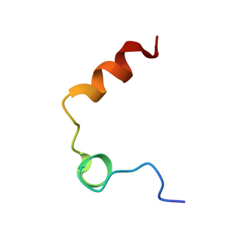
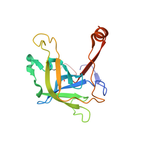
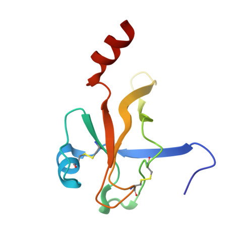
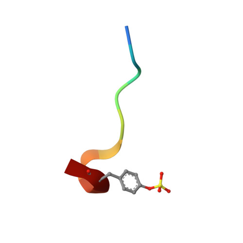
    3PO1

  • PubMed Abstract: 

    In a project to find novel neutral P1 fragments for the synthesis of thrombin inhibitors with improved pharmacokinetic properties, fragments containing a benzothiazole guanidine scaffold were identified as weak thrombin inhibitors. WaterLOGSY (Water-Ligand Observed via Gradient SpectroscopY) NMR was used to detect fragments binding to thrombin and these fragments were followed up by Biacore A100 affinity measurements and enzyme assays. A crystal structure of the most potent compound with thrombin was obtained and revealed an unexpected binding mode as well as the key interactions of the fragment with the protein. Based on these results, the structure-based design and synthesis of a small series of optimized novel substituted benzothiazole guanidines with comparatively low pK(a) values was accomplished. Testing of these compounds against human trypsin I and human trypsin IV revealed unexpected inhibitory activity and selectivity of some of the compounds, making them attractive starting points for selective trypsin inhibitors.


  • Organizational Affiliation

    AstraZeneca R&D Mölndal, SE-43183 Mölndal, Sweden. Michael.Karle@astrazeneca.com


Macromolecules
Find similar proteins by:  (by identity cutoff)  |  3D Structure
Entity ID: 1
MoleculeChains Sequence LengthOrganismDetailsImage
Thrombin light chain27Homo sapiensMutation(s): 0 
EC: 3.4.21.5
UniProt & NIH Common Fund Data Resources
Find proteins for P00734 (Homo sapiens)
Explore P00734 
Go to UniProtKB:  P00734
PHAROS:  P00734
GTEx:  ENSG00000180210 
Entity Groups  
Sequence Clusters30% Identity50% Identity70% Identity90% Identity95% Identity100% Identity
UniProt GroupP00734
Sequence Annotations
Expand
  • Reference Sequence
Find similar proteins by:  (by identity cutoff)  |  3D Structure
Entity ID: 2
MoleculeChains Sequence LengthOrganismDetailsImage
Thrombin heavy chain147Homo sapiensMutation(s): 0 
EC: 3.4.21.5
UniProt & NIH Common Fund Data Resources
Find proteins for P00734 (Homo sapiens)
Explore P00734 
Go to UniProtKB:  P00734
PHAROS:  P00734
GTEx:  ENSG00000180210 
Entity Groups  
Sequence Clusters30% Identity50% Identity70% Identity90% Identity95% Identity100% Identity
UniProt GroupP00734
Sequence Annotations
Expand
  • Reference Sequence
Find similar proteins by:  (by identity cutoff)  |  3D Structure
Entity ID: 3
MoleculeChains Sequence LengthOrganismDetailsImage
Thrombin heavy chain102Homo sapiensMutation(s): 0 
EC: 3.4.21.5
UniProt & NIH Common Fund Data Resources
Find proteins for P00734 (Homo sapiens)
Explore P00734 
Go to UniProtKB:  P00734
PHAROS:  P00734
GTEx:  ENSG00000180210 
Entity Groups  
Sequence Clusters30% Identity50% Identity70% Identity90% Identity95% Identity100% Identity
UniProt GroupP00734
Sequence Annotations
Expand
  • Reference Sequence

Find similar proteins by:  Sequence   |   3D Structure  

Entity ID: 4
MoleculeChains Sequence LengthOrganismDetailsImage
thrombin peptide10Homo sapiensMutation(s): 0 
Entity Groups  
Sequence Clusters30% Identity50% Identity70% Identity90% Identity95% Identity100% Identity
Sequence Annotations
Expand
  • Reference Sequence
Small Molecules
Ligands 3 Unique
IDChains Name / Formula / InChI Key2D Diagram3D Interactions
MKY
Query on MKY

Download Ideal Coordinates CCD File 
E [auth C]ethyl [(2Z)-2-(carbamimidoylimino)-6-hydroxy-1,3-benzothiazol-3(2H)-yl]acetate
C12 H14 N4 O3 S
QHEKEKSMRXTAKP-QINSGFPZSA-N
ACT
Query on ACT

Download Ideal Coordinates CCD File 
G [auth C]ACETATE ION
C2 H3 O2
QTBSBXVTEAMEQO-UHFFFAOYSA-M
NA
Query on NA

Download Ideal Coordinates CCD File 
F [auth C]SODIUM ION
Na
FKNQFGJONOIPTF-UHFFFAOYSA-N
Modified Residues  1 Unique
IDChains TypeFormula2D DiagramParent
TYS
Query on TYS
D
L-PEPTIDE LINKINGC9 H11 N O6 STYR
Binding Affinity Annotations 
IDSourceBinding Affinity
MKY PDBBind:  3PO1 Kd: 9.50e+4 (nM) from 1 assay(s)
Binding MOAD:  3PO1 Kd: 9.50e+4 (nM) from 1 assay(s)
Experimental Data & Validation

Experimental Data

  • Method: X-RAY DIFFRACTION
  • Resolution: 1.65 Å
  • R-Value Free: 0.260 
  • R-Value Work: 0.227 
  • R-Value Observed: 0.229 
  • Space Group: C 1 2 1
Unit Cell:
Length ( Å )Angle ( ˚ )
a = 68.662α = 90
b = 71.414β = 99.82
c = 71.88γ = 90
Software Package:
Software NamePurpose
MOSFLMdata reduction
SCALAdata scaling
REFMACrefinement
PDB_EXTRACTdata extraction

Structure Validation

View Full Validation Report


Ligand Structure Quality Assessment 


Entry History 

Deposition Data

  • Released Date: 2011-11-23 
  • Deposition Author(s): Xue, Y.

Revision History  (Full details and data files)

  • Version 1.0: 2011-11-23
    Type: Initial release
  • Version 1.1: 2012-07-25
    Changes: Database references