3NMN

Crystal structure of pyrabactin-bound abscisic acid receptor PYL1 in complex with type 2C protein phosphatase ABI1


Experimental Data Snapshot

  • Method: X-RAY DIFFRACTION
  • Resolution: 2.15 Å
  • R-Value Free: 0.248 
  • R-Value Work: 0.210 
  • R-Value Observed: 0.213 

wwPDB Validation   3D Report Full Report


Ligand Structure Quality Assessment 


This is version 1.2 of the entry. See complete history


Literature

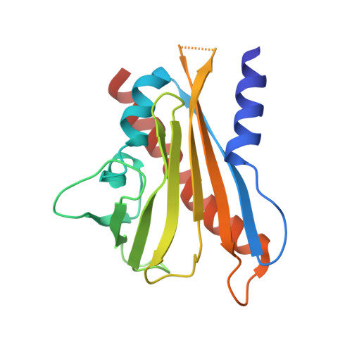
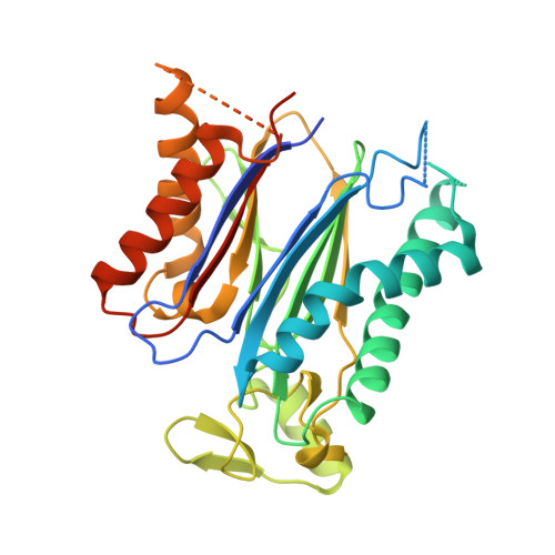
Identification and mechanism of ABA receptor antagonism.

Melcher, K.Xu, Y.Ng, L.M.Zhou, X.E.Soon, F.F.Chinnusamy, V.Suino-Powell, K.M.Kovach, A.Tham, F.S.Cutler, S.R.Li, J.Yong, E.L.Zhu, J.K.Xu, H.E.

(2010) Nat Struct Mol Biol 17: 1102-1108

  • DOI: https://doi.org/10.1038/nsmb.1887
  • Primary Citation of Related Structures:  
    3NMH, 3NMN, 3NMP, 3NMT, 3NMV

  • PubMed Abstract: 

    The phytohormone abscisic acid (ABA) functions through a family of fourteen PYR/PYL receptors, which were identified by resistance to pyrabactin, a synthetic inhibitor of seed germination. ABA activates these receptors to inhibit type 2C protein phosphatases, such as ABI1, yet it remains unclear whether these receptors can be antagonized. Here we demonstrate that pyrabactin is an agonist of PYR1 and PYL1 but is unexpectedly an antagonist of PYL2. Crystal structures of the PYL2-pyrabactin and PYL1-pyrabactin-ABI1 complexes reveal the mechanism responsible for receptor-selective activation and inhibition, which enables us to design mutations that convert PYL1 to a pyrabactin-inhibited receptor and PYL2 to a pyrabactin-activated receptor and to identify new pyrabactin-based ABA receptor agonists. Together, our results establish a new concept of ABA receptor antagonism, illustrate its underlying mechanisms and provide a rational framework for discovering novel ABA receptor ligands.


  • Organizational Affiliation

    Laboratory of Structural Sciences, Van Andel Research Institute, Grand Rapids, Michigan, USA. Karsten.Melcher@vai.org


Macromolecules
Find similar proteins by:  (by identity cutoff)  |  3D Structure
Entity ID: 1
MoleculeChains Sequence LengthOrganismDetailsImage
Abscisic acid receptor PYL1
A, C
178Arabidopsis thalianaMutation(s): 0 
Gene Names: PYL1RCAR12At5g46790MZA15.21
UniProt
Find proteins for Q8VZS8 (Arabidopsis thaliana)
Explore Q8VZS8 
Go to UniProtKB:  Q8VZS8
Entity Groups  
Sequence Clusters30% Identity50% Identity70% Identity90% Identity95% Identity100% Identity
UniProt GroupQ8VZS8
Sequence Annotations
Expand
  • Reference Sequence
Find similar proteins by:  (by identity cutoff)  |  3D Structure
Entity ID: 2
MoleculeChains Sequence LengthOrganismDetailsImage
Protein phosphatase 2C 56
B, D
319Arabidopsis thalianaMutation(s): 0 
Gene Names: ABI1At4g26080F20B18.190
EC: 3.1.3.16
UniProt
Find proteins for P49597 (Arabidopsis thaliana)
Explore P49597 
Go to UniProtKB:  P49597
Entity Groups  
Sequence Clusters30% Identity50% Identity70% Identity90% Identity95% Identity100% Identity
UniProt GroupP49597
Sequence Annotations
Expand
  • Reference Sequence
Experimental Data & Validation

Experimental Data

  • Method: X-RAY DIFFRACTION
  • Resolution: 2.15 Å
  • R-Value Free: 0.248 
  • R-Value Work: 0.210 
  • R-Value Observed: 0.213 
  • Space Group: P 1
Unit Cell:
Length ( Å )Angle ( ˚ )
a = 59.978α = 115.78
b = 66.71β = 95.43
c = 72.598γ = 105.6
Software Package:
Software NamePurpose
HKL-2000data collection
PHASERphasing
REFMACrefinement
DENZOdata reduction
SCALEPACKdata scaling

Structure Validation

View Full Validation Report


Ligand Structure Quality Assessment 


Entry History 

Deposition Data

Revision History  (Full details and data files)

  • Version 1.0: 2010-08-25
    Type: Initial release
  • Version 1.1: 2011-07-13
    Changes: Version format compliance
  • Version 1.2: 2023-09-06
    Changes: Data collection, Database references, Derived calculations, Refinement description