The role of Zn2+ on the structure and stability of murine adenosine deaminase.
Niu, W., Shu, Q., Chen, Z., Mathews, S., Di Cera, E., Frieden, C.(2010) J Phys Chem B 114: 16156-16165
- PubMed: 20815357
- DOI: https://doi.org/10.1021/jp106041v
- Primary Citation of Related Structures:
3MVI, 3MVT - PubMed Abstract:
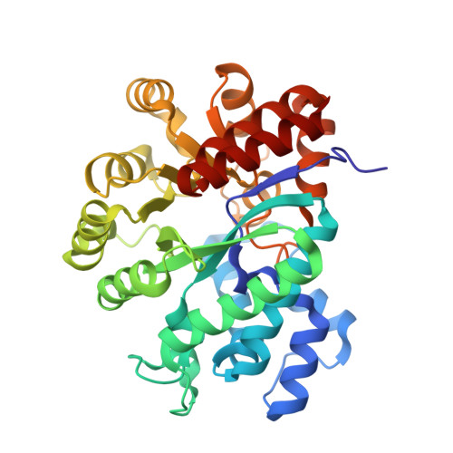
Adenosine deaminase (ADA) is a key enzyme in purine metabolism and crucial for normal immune competence. It is a 40 kDa monomeric TIM-barrel protein containing a tightly bound Zn(2+), which is required for activity. In this study, we have investigated the role of Zn(2+) with respect to ADA structure and stability. After removing Zn(2+), the crystallographic structure of the protein remains highly ordered and similar to that of the holo protein with structural changes limited to regions capping the active site pocket. The stability of the protein, however, is decreased significantly in the absence of Zn(2+). Denaturation with urea shows the midpoint to be about 3.5 M for the apo enzyme, compared with 6.4 M for the holo enzyme. ADA contains four tryptophan residues distant from the Zn(2+) site. (19)F NMR studies in the presence and absence of Zn(2+) were carried out after incorporation of 6-(19)F-tryptophan. Chemical shift differences were observed for three of the four tryptophan residues, suggesting that, in contrast to the X-ray data, Zn(2+)-induced structural changes are propagated throughout the protein. Changes throughout the structure as suggested by the NMR data may explain the lower stability of the Zn(2+)-free protein. Real-time (19)F NMR spectroscopy measuring the loss of Zn(2+) showed that structural changes correlated with the loss of enzymatic activity.
Organizational Affiliation:
Edward A. Doisy Department of Biochemistry and Molecular Biology, Saint Louis University School of Medicine, St. Louis, Missouri 63104, USA.