


Crystal structure of deoxyuridine 5-triphosphate nucleotidohydrolase from Entamoeba histolytica
Abendroth, J., Sankaran, B., Arakaki, T., Staker, B.To be published.
Experimental Data Snapshot
wwPDB Validation 3D Report Full Report

Entity ID: 1 | |||||
|---|---|---|---|---|---|
| Molecule | Chains | Sequence Length | Organism | Details | Image |
| Deoxyuridine 5'-triphosphate nucleotidohydrolase | 163 | Entamoeba histolytica HM-1:IMSS | Mutation(s): 0 Gene Names: EHI_189420 | 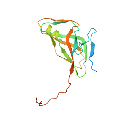 | |
UniProt | |||||
Find proteins for C4M4M6 (Entamoeba histolytica (strain ATCC 30459 / HM-1:IMSS / ABRM)) Explore C4M4M6 Go to UniProtKB: C4M4M6 | |||||
Entity Groups | |||||
| Sequence Clusters | 30% Identity50% Identity70% Identity90% Identity95% Identity100% Identity | ||||
| UniProt Group | C4M4M6 | ||||
Sequence AnnotationsExpand | |||||
| |||||
| Ligands 2 Unique | |||||
|---|---|---|---|---|---|
| ID | Chains | Name / Formula / InChI Key | 2D Diagram | 3D Interactions | |
| GOL Query on GOL | C [auth A] | GLYCEROL C3 H8 O3 PEDCQBHIVMGVHV-UHFFFAOYSA-N |  | ||
| EDO Query on EDO | B [auth A] | 1,2-ETHANEDIOL C2 H6 O2 LYCAIKOWRPUZTN-UHFFFAOYSA-N |  | ||
| Length ( Å ) | Angle ( ˚ ) |
|---|---|
| a = 63.14 | α = 90 |
| b = 63.14 | β = 90 |
| c = 108.37 | γ = 120 |
| Software Name | Purpose |
|---|---|
| BOS | data collection |
| PHASER | phasing |
| REFMAC | refinement |
| XDS | data reduction |
| XSCALE | data scaling |
RCSB PDB (citation) is hosted by
RCSB PDB is a member of the

