3KW0

Crystal structure of Cysteine peptidase (NP_982244.1) from BACILLUS CEREUS ATCC 10987 at 2.50 A resolution


Experimental Data Snapshot

  • Method: X-RAY DIFFRACTION
  • Resolution: 2.50 Å
  • R-Value Free: 0.219 
  • R-Value Work: 0.192 
  • R-Value Observed: 0.193 

wwPDB Validation   3D Report Full Report


This is version 1.5 of the entry. See complete history


Literature

Structural Analysis of Papain-Like NlpC/P60 Superfamily Enzymes with a Circularly Permuted Topology Reveals Potential Lipid Binding Sites.

Xu, Q.Rawlings, N.D.Chiu, H.J.Jaroszewski, L.Klock, H.E.Knuth, M.W.Miller, M.D.Elsliger, M.A.Deacon, A.M.Godzik, A.Lesley, S.A.Wilson, I.A.

(2011) PLoS One 6: e22013-e22013

  • DOI: https://doi.org/10.1371/journal.pone.0022013
  • Primary Citation of Related Structures:  
    3KW0

  • PubMed Abstract: 

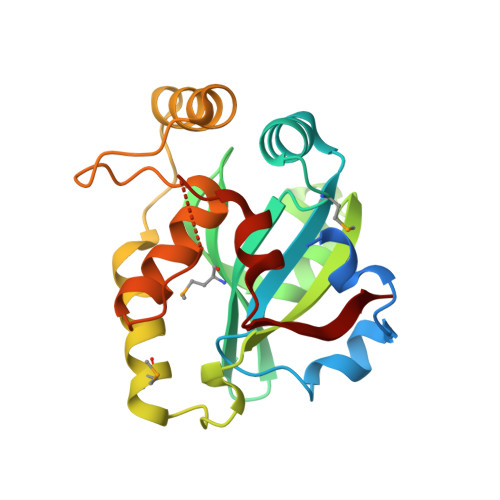
    NlpC/P60 superfamily papain-like enzymes play important roles in all kingdoms of life. Two members of this superfamily, LRAT-like and YaeF/YiiX-like families, were predicted to contain a catalytic domain that is circularly permuted such that the catalytic cysteine is located near the C-terminus, instead of at the N-terminus. These permuted enzymes are widespread in virus, pathogenic bacteria, and eukaryotes. We determined the crystal structure of a member of the YaeF/YiiX-like family from Bacillus cereus in complex with lysine. The structure, which adopts a ligand-induced, "closed" conformation, confirms the circular permutation of catalytic residues. A comparative analysis of other related protein structures within the NlpC/P60 superfamily is presented. Permutated NlpC/P60 enzymes contain a similar conserved core and arrangement of catalytic residues, including a Cys/His-containing triad and an additional conserved tyrosine. More surprisingly, permuted enzymes have a hydrophobic S1 binding pocket that is distinct from previously characterized enzymes in the family, indicative of novel substrate specificity. Further analysis of a structural homolog, YiiX (PDB 2if6) identified a fatty acid in the conserved hydrophobic pocket, thus providing additional insights into possible function of these novel enzymes.


  • Organizational Affiliation

    Joint Center for Structural Genomics, La Jolla, California, United States of America.


Macromolecules
Find similar proteins by:  (by identity cutoff)  |  3D Structure
Entity ID: 1
MoleculeChains Sequence LengthOrganismDetailsImage
Cysteine peptidase
A, B, C, D
214Bacillus cereus ATCC 10987Mutation(s): 0 
Gene Names: BCE_A0238
UniProt
Find proteins for Q74NK7 (Bacillus cereus (strain ATCC 10987 / NRS 248))
Explore Q74NK7 
Go to UniProtKB:  Q74NK7
Entity Groups  
Sequence Clusters30% Identity50% Identity70% Identity90% Identity95% Identity100% Identity
UniProt GroupQ74NK7
Sequence Annotations
Expand
  • Reference Sequence
Small Molecules
Ligands 2 Unique
IDChains Name / Formula / InChI Key2D Diagram3D Interactions
LYS
Query on LYS

Download Ideal Coordinates CCD File 
E [auth A],
I [auth B],
M [auth C],
P [auth D]
LYSINE
C6 H15 N2 O2
KDXKERNSBIXSRK-YFKPBYRVSA-O
CL
Query on CL

Download Ideal Coordinates CCD File 
F [auth A]
G [auth A]
H [auth A]
J [auth B]
K [auth B]
F [auth A],
G [auth A],
H [auth A],
J [auth B],
K [auth B],
L [auth B],
N [auth C],
O [auth C],
Q [auth D]
CHLORIDE ION
Cl
VEXZGXHMUGYJMC-UHFFFAOYSA-M
Modified Residues  1 Unique
IDChains TypeFormula2D DiagramParent
MSE
Query on MSE
A, B, C, D
L-PEPTIDE LINKINGC5 H11 N O2 SeMET
Experimental Data & Validation

Experimental Data

  • Method: X-RAY DIFFRACTION
  • Resolution: 2.50 Å
  • R-Value Free: 0.219 
  • R-Value Work: 0.192 
  • R-Value Observed: 0.193 
  • Space Group: P 65
  • Diffraction Data: https://doi.org/10.18430/M33KW0
Unit Cell:
Length ( Å )Angle ( ˚ )
a = 64.96α = 90
b = 64.96β = 90
c = 407.8γ = 120
Software Package:
Software NamePurpose
REFMACrefinement
PHENIXrefinement
SHELXphasing
MolProbitymodel building
XSCALEdata scaling
PDB_EXTRACTdata extraction
XDSdata reduction
SHELXDphasing
autoSHARPphasing

Structure Validation

View Full Validation Report


Entry History 

Deposition Data

Revision History  (Full details and data files)

  • Version 1.0: 2009-12-15
    Type: Initial release
  • Version 1.1: 2011-07-13
    Changes: Advisory, Source and taxonomy, Version format compliance
  • Version 1.2: 2011-08-10
    Changes: Database references
  • Version 1.3: 2017-11-01
    Changes: Refinement description
  • Version 1.4: 2019-07-17
    Changes: Data collection, Derived calculations, Refinement description
  • Version 1.5: 2023-02-01
    Changes: Database references, Derived calculations