3JUR

The crystal structure of a hyperthermoactive Exopolygalacturonase from Thermotoga maritima


Experimental Data Snapshot

  • Method: X-RAY DIFFRACTION
  • Resolution: 2.05 Å
  • R-Value Free: 0.220 
  • R-Value Work: 0.175 
  • R-Value Observed: 0.177 

wwPDB Validation   3D Report Full Report


This is version 1.2 of the entry. See complete history


Literature

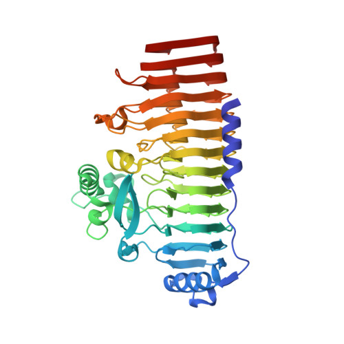
The crystal structure of a hyperthermoactive exopolygalacturonase from Thermotoga maritima reveals a unique tetramer

Pijning, T.van Pouderoyen, G.Kluskens, L.van der Oost, J.Dijkstra, B.W.

(2009) FEBS Lett 

  • DOI: https://doi.org/10.1016/j.febslet.2009.10.047
  • Primary Citation of Related Structures:  
    3JUR

  • PubMed Abstract: 

    The exopolygalacturonase from Thermotoga maritima is the most thermoactive and thermostable pectinase known to date. Here we present its crystal structure at 2.05A resolution. High structural homology around the active site allowed us to propose a model for substrate binding, explaining the exo-cleavage activity and specificity for non-methylated saturated galacturonate at the non-reducing end. Furthermore, the structure reveals unique features that contribute to the formation of stable tetramers in solution. Such an oligomerization has not been observed before for polygalacturonases.


  • Organizational Affiliation

    Laboratory of Biophysical Chemistry, University of Groningen, Nijenborgh 4, 9747 AG Groningen, The Netherlands.


Macromolecules
Find similar proteins by:  (by identity cutoff)  |  3D Structure
Entity ID: 1
MoleculeChains Sequence LengthOrganismDetailsImage
Exo-poly-alpha-D-galacturonosidase
A, B, C, D
448Thermotoga maritimaMutation(s): 0 
Gene Names: TM0437
EC: 3.2.1.15
UniProt
Find proteins for Q9WYR8 (Thermotoga maritima (strain ATCC 43589 / DSM 3109 / JCM 10099 / NBRC 100826 / MSB8))
Explore Q9WYR8 
Go to UniProtKB:  Q9WYR8
Entity Groups  
Sequence Clusters30% Identity50% Identity70% Identity90% Identity95% Identity100% Identity
UniProt GroupQ9WYR8
Sequence Annotations
Expand
  • Reference Sequence
Experimental Data & Validation

Experimental Data

  • Method: X-RAY DIFFRACTION
  • Resolution: 2.05 Å
  • R-Value Free: 0.220 
  • R-Value Work: 0.175 
  • R-Value Observed: 0.177 
  • Space Group: P 21 21 21
Unit Cell:
Length ( Å )Angle ( ˚ )
a = 63.274α = 90
b = 155.904β = 90
c = 200.652γ = 90
Software Package:
Software NamePurpose
PHASERphasing
REFMACrefinement
XDSdata reduction
SCALAdata scaling

Structure Validation

View Full Validation Report


Entry History 

Deposition Data

Revision History  (Full details and data files)

  • Version 1.0: 2009-11-17
    Type: Initial release
  • Version 1.1: 2011-07-13
    Changes: Advisory, Version format compliance
  • Version 1.2: 2023-11-01
    Changes: Data collection, Database references, Refinement description