3IAG

CSL (RBP-Jk) bound to HES-1 nonconsensus site


Experimental Data Snapshot

  • Method: X-RAY DIFFRACTION
  • Resolution: 2.00 Å
  • R-Value Free: 0.243 
  • R-Value Work: 0.197 
  • R-Value Observed: 0.199 

wwPDB Validation   3D Report Full Report


Ligand Structure Quality Assessment 


This is version 1.4 of the entry. See complete history


Literature

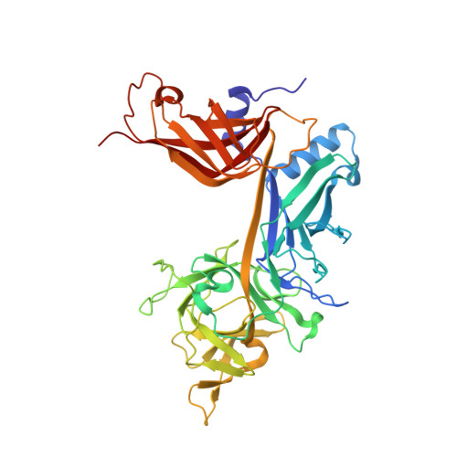
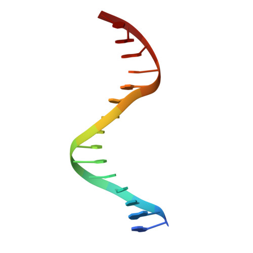
Thermodynamic and structural insights into CSL-DNA complexes.

Friedmann, D.R.Kovall, R.A.

(2010) Protein Sci 19: 34-46

  • DOI: https://doi.org/10.1002/pro.280
  • Primary Citation of Related Structures:  
    3IAG

  • PubMed Abstract: 

    The Notch pathway is an intercellular signaling mechanism that plays important roles in cell fates decisions throughout the developing and adult organism. Extracellular complexation of Notch receptors with ligands ultimately results in changes in gene expression, which is regulated by the nuclear effector of the pathway, CSL (C-promoter binding factor 1 (CBF-1), suppressor of hairless (Su(H)), lin-12 and glp-1 (Lag-1)). CSL is a DNA binding protein that is involved in both repression and activation of transcription from genes that are responsive to Notch signaling. One well-characterized Notch target gene is hairy and enhancer of split-1 (HES-1), which is regulated by a promoter element consisting of two CSL binding sites oriented in a head-to-head arrangement. Although previous studies have identified in vivo and consensus binding sites for CSL, and crystal structures of these complexes have been determined, to date, a quantitative description of the energetics that underlie CSL-DNA binding is unknown. Here, we provide a thermodynamic and structural analysis of the interaction between CSL and the two individual sites that comprise the HES-1 promoter element. Our comprehensive studies that analyze binding as a function of temperature, salt, and pH reveal moderate, but distinct, differences in the affinities of CSL for the two HES-1 binding sites. Similarly, our structural results indicate that overall CSL binds both DNA sites in a similar manner; however, minor changes are observed in both the conformation of CSL and DNA. Taken together, our results provide a quantitative and biophysical basis for understanding how CSL interacts with DNA sites in vivo.


  • Organizational Affiliation

    Department of Molecular Genetics, Biochemistry and Microbiology, University of Cincinnati College of Medicine, Cincinnati, Ohio 45267-0524, USA.


Macromolecules

Find similar proteins by:  (by identity cutoff)  |  3D Structure
Entity ID: 3
MoleculeChains Sequence LengthOrganismDetailsImage
Recombining binding protein suppressor of hairless422Mus musculusMutation(s): 0 
Gene Names: Igkjrb1IgkrsbpRBP-JkRbpjRbpsuh
UniProt & NIH Common Fund Data Resources
Find proteins for P31266 (Mus musculus)
Explore P31266 
Go to UniProtKB:  P31266
IMPC:  MGI:96522
Entity Groups  
Sequence Clusters30% Identity50% Identity70% Identity90% Identity95% Identity100% Identity
UniProt GroupP31266
Sequence Annotations
Expand
  • Reference Sequence

Find similar nucleic acids by:  Sequence   |   3D Structure  

Entity ID: 1
MoleculeChains LengthOrganismImage
5'-D(*AP*AP*TP*CP*TP*TP*TP*CP*AP*CP*AP*CP*GP*AP*T)-3'15N/A
Sequence Annotations
Expand
  • Reference Sequence

Find similar nucleic acids by:  Sequence   |   3D Structure  

Entity ID: 2
MoleculeChains LengthOrganismImage
5'-D(*TP*TP*AP*TP*CP*GP*TP*GP*TP*GP*AP*AP*AP*GP*A)-3'15N/A
Sequence Annotations
Expand
  • Reference Sequence
Experimental Data & Validation

Experimental Data

  • Method: X-RAY DIFFRACTION
  • Resolution: 2.00 Å
  • R-Value Free: 0.243 
  • R-Value Work: 0.197 
  • R-Value Observed: 0.199 
  • Space Group: P 21 21 2
Unit Cell:
Length ( Å )Angle ( ˚ )
a = 63.451α = 90
b = 93.137β = 90
c = 112.569γ = 90
Software Package:
Software NamePurpose
DENZOdata reduction
SCALEPACKdata scaling
REFMACrefinement
PDB_EXTRACTdata extraction

Structure Validation

View Full Validation Report


Ligand Structure Quality Assessment 


Entry History 

Deposition Data

Revision History  (Full details and data files)

  • Version 1.0: 2009-11-10
    Type: Initial release
  • Version 1.1: 2011-07-13
    Changes: Version format compliance
  • Version 1.2: 2017-11-01
    Changes: Refinement description
  • Version 1.3: 2020-07-29
    Type: Remediation
    Reason: Carbohydrate remediation
    Changes: Derived calculations, Structure summary
  • Version 1.4: 2024-02-21
    Changes: Data collection, Database references, Structure summary