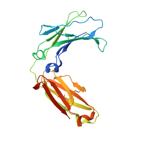
Structure of the murine unglycosylated IgG1 Fc fragment
Feige, M.J., Nath, S., Catharino, S.R., Weinfurtner, D., Steinbacher, S., Buchner, J.(2009) J Mol Biol 391: 599-608
- PubMed: 19559712
- DOI: https://doi.org/10.1016/j.jmb.2009.06.048
- Primary Citation of Related Structures:
3HKF - PubMed Abstract:
A prototypic IgG antibody can be divided into two major structural units: the antigen-binding fragment (Fab) and the Fc fragment that mediates effector functions. The IgG Fc fragment is a homodimer of the two C-terminal domains (C(H)2 and C(H)3) of the heavy chains. Characteristic of the Fc part is the presence of a sugar moiety at the inner face of the C(H)2 domains. The structure of this complex branched oligosaccharide is generally resolved in crystal structures of Fc fragments due to numerous well-defined sugar-protein interactions and a small number of sugar-sugar interactions. This suggested that sugars play an important role in the structure of the Fc fragment. To address this question directly, we determined the crystal structure of the unglycosylated Fc fragment of the murine IgG1 MAK33. The structures of the C(H)3 domains of the unglycosylated Fc fragment superimpose perfectly with the structure of the isolated MAK33 C(H)3 domain. The unglycosylated C(H)2 domains, in contrast, approach each other much more closely compared to known structures of partly deglycosylated Fc fragments with rigid-body motions between 10 and 14 A, leading to a strongly "closed" conformation of the unglycosylated Fc fragment. The glycosylation sites in the C'E loop and the BC and FG loops are well defined in the unglycosylated C(H)2 domain, however, with increased mobility and with a significant displacement of about 4.9 A for the unglycosylated Asn residue compared to the glycosylated structure. Thus, glycosylation both stabilizes the C'E-loop conformation within the C(H)2 domain and also helps to ensure an "open" conformation, as seen upon Fc receptor binding. These structural data provide a rationale for the observation that deglycosylation of antibodies often compromises their ability to bind and activate Fcgamma receptors.
Organizational Affiliation:
Department Chemie and Center for Integrated Protein Science Munich, Technische Universität München, 85747 Garching, Germany.