3DUU

Crystal structure of SAG506-01, orthorhombic, twinned, crystal 2


Experimental Data Snapshot

  • Method: X-RAY DIFFRACTION
  • Resolution: 1.95 Å
  • R-Value Free: 0.217 
  • R-Value Work: 0.194 
  • R-Value Observed: 0.195 

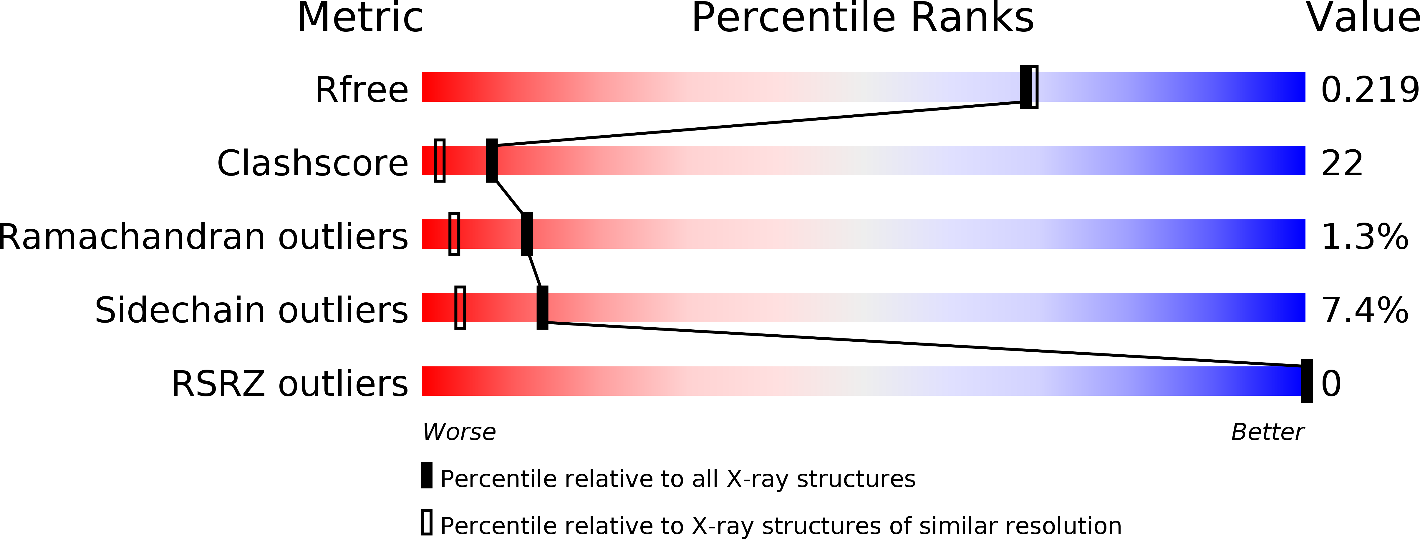
wwPDB Validation   3D Report Full Report


Ligand Structure Quality Assessment 


This is version 1.2 of the entry. See complete history


Literature

Pseudo-symmetry and twinning in crystals of homologous antibody Fv fragments.

Brooks, C.L.Blackler, R.J.Gerstenbruch, S.Kosma, P.Muller-Loennies, S.Brade, H.Evans, S.V.

(2008) Acta Crystallogr D Biol Crystallogr 64: 1250-1258

  • DOI: https://doi.org/10.1107/S0907444908033453
  • Primary Citation of Related Structures:  
    3DUR, 3DUS, 3DUU, 3DV4, 3DV6

  • PubMed Abstract: 

    A difference of seven conservative amino-acid substitutions between two single-chain antibodies (scFvs) specific for chlamydial lipopolysaccharide does not significantly affect their molecular structures or packing contacts, but dramatically affects their crystallization. The structure of the variable domain (Fv) of SAG173-04 was solved to 1.86 A resolution and an R(cryst) of 18.9% in space group P2(1)2(1)2(1). Crystals of the homologous SAG506-01 diffracted to 1.95 A resolution and appeared at first to have Patterson symmetry I4/m or P4/mmm; however, no solution could be found in space groups belonging to the former and refinement in the only solution corresponding to the latter (in space group P4(3)2(1)2) stalled at R(free) = 30.0%. Detailed examination of the diffraction data revealed that the crystal was likely to be twinned and that the correct space group was P2(1)2(1)2(1). Both translational pseudo-symmetry and pseudo-merohedral twinning were observed in one crystal of SAG506-01 and pseudo-merohedral twinning was observed for a second crystal. The final R factor for SAG506-01 after refinement in P2(1)2(1)2(1) was 20.5%.


  • Organizational Affiliation

    University of Victoria, Department of Biochemistry and Microbiology, Victoria BC V8P 3P6, Canada.


Macromolecules
Find similar proteins by:  (by identity cutoff)  |  3D Structure
Entity ID: 1
MoleculeChains Sequence LengthOrganismDetailsImage
antibody Fv fragment SAG506-01
A, C
112Mus musculusMutation(s): 0 
UniProt
Find proteins for A0N262 (Mus musculus)
Explore A0N262 
Go to UniProtKB:  A0N262
Entity Groups  
Sequence Clusters30% Identity50% Identity70% Identity90% Identity95% Identity100% Identity
UniProt GroupA0N262
Sequence Annotations
Expand
  • Reference Sequence
Find similar proteins by:  (by identity cutoff)  |  3D Structure
Entity ID: 2
MoleculeChains Sequence LengthOrganismDetailsImage
Ig-like protein
B, D
121Mus musculusMutation(s): 0 
UniProt
Find proteins for A2NU21 (Mus musculus)
Explore A2NU21 
Go to UniProtKB:  A2NU21
Entity Groups  
Sequence Clusters30% Identity50% Identity70% Identity90% Identity95% Identity100% Identity
UniProt GroupA2NU21
Sequence Annotations
Expand
  • Reference Sequence
Experimental Data & Validation

Experimental Data

  • Method: X-RAY DIFFRACTION
  • Resolution: 1.95 Å
  • R-Value Free: 0.217 
  • R-Value Work: 0.194 
  • R-Value Observed: 0.195 
  • Space Group: P 21 21 21
Unit Cell:
Length ( Å )Angle ( ˚ )
a = 71.6α = 90
b = 71.76β = 90
c = 86.21γ = 90
Software Package:
Software NamePurpose
d*TREKdata scaling
PHASERphasing
PHENIXrefinement
PDB_EXTRACTdata extraction
d*TREKdata reduction

Structure Validation

View Full Validation Report


Ligand Structure Quality Assessment 


Entry History 

Deposition Data

Revision History  (Full details and data files)

  • Version 1.0: 2008-12-02
    Type: Initial release
  • Version 1.1: 2011-07-13
    Changes: Version format compliance
  • Version 1.2: 2020-07-29
    Type: Remediation
    Reason: Carbohydrate remediation
    Changes: Data collection, Derived calculations, Structure summary