3DJF

Crystal Structure of Schistosoma mansoni Purine Nucleoside Phosphorylase in a complex with BCX-34


Experimental Data Snapshot

  • Method: X-RAY DIFFRACTION
  • Resolution: 2.30 Å
  • R-Value Free: 0.259 
  • R-Value Work: 0.201 
  • R-Value Observed: 0.204 

wwPDB Validation   3D Report Full Report


Ligand Structure Quality Assessment 


This is version 1.2 of the entry. See complete history


Literature

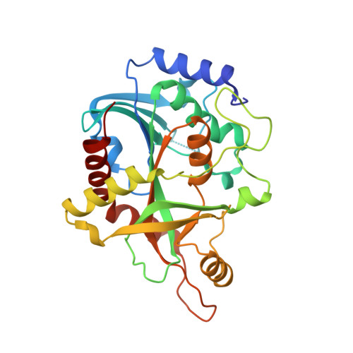
Structural basis for selective inhibition of purine nucleoside phosphorylase from Schistosoma mansoni: kinetic and structural studies.

Castilho, M.S.Postigo, M.P.Pereira, H.M.Oliva, G.Andricopulo, A.D.

(2010) Bioorg Med Chem 18: 1421-1427

  • DOI: https://doi.org/10.1016/j.bmc.2010.01.022
  • Primary Citation of Related Structures:  
    3DJF, 3IEX

  • PubMed Abstract: 

    Selectivity plays a crucial role in the design of enzyme inhibitors as novel antiparasitic agents, particularly in cases where the target enzyme is also present in the human host. Purine nucleoside phosphorylase from Schistosoma mansoni (SmPNP) is an attractive target for the discovery of potential antischistosomal agents. In the present work, kinetic studies were carried out in order to determine the inhibitory potency, mode of action and enzyme selectivity of a series of inhibitors of SmPNP. In addition, crystallographic studies provided important structural insights for rational inhibitor design, revealing consistent structural differences in the binding mode of the inhibitors in the active sites of the SmPNP and human PNP (HsPNP) structures. The molecular information gathered in this work should be useful for future medicinal chemistry efforts in the design of new inhibitors of SmPNP having increased affinity and selectivity.


  • Organizational Affiliation

    Universidade Federal da Bahia, Faculdade de Farmácia, R. Barão de Jeremoabo, Salvador-BA, Brazil. castilho@ufba.br


Macromolecules
Find similar proteins by:  (by identity cutoff)  |  3D Structure
Entity ID: 1
MoleculeChains Sequence LengthOrganismDetailsImage
Purine-nucleoside phosphorylase
A, B, C
287Schistosoma mansoniMutation(s): 0 
EC: 2.4.2.1
UniProt
Find proteins for Q9BMI9 (Schistosoma mansoni)
Explore Q9BMI9 
Go to UniProtKB:  Q9BMI9
Entity Groups  
Sequence Clusters30% Identity50% Identity70% Identity90% Identity95% Identity100% Identity
UniProt GroupQ9BMI9
Sequence Annotations
Expand
  • Reference Sequence
Small Molecules
Ligands 3 Unique
IDChains Name / Formula / InChI Key2D Diagram3D Interactions
BC3
Query on BC3

Download Ideal Coordinates CCD File 
F [auth A],
H [auth B],
K [auth C]
2-amino-7-(pyridin-3-ylmethyl)-3,5-dihydro-4H-pyrrolo[3,2-d]pyrimidin-4-one
C12 H11 N5 O
DOHVAKFYAHLCJP-UHFFFAOYSA-N
SO4
Query on SO4

Download Ideal Coordinates CCD File 
D [auth A],
I [auth C]
SULFATE ION
O4 S
QAOWNCQODCNURD-UHFFFAOYSA-L
DMS
Query on DMS

Download Ideal Coordinates CCD File 
E [auth A],
G [auth B],
J [auth C]
DIMETHYL SULFOXIDE
C2 H6 O S
IAZDPXIOMUYVGZ-UHFFFAOYSA-N
Binding Affinity Annotations 
IDSourceBinding Affinity
BC3 Binding MOAD:  3DJF IC50: 800 (nM) from 1 assay(s)
PDBBind:  3DJF IC50: 800 (nM) from 1 assay(s)
Experimental Data & Validation

Experimental Data

  • Method: X-RAY DIFFRACTION
  • Resolution: 2.30 Å
  • R-Value Free: 0.259 
  • R-Value Work: 0.201 
  • R-Value Observed: 0.204 
  • Space Group: P 21 21 21
Unit Cell:
Length ( Å )Angle ( ˚ )
a = 49.577α = 90
b = 132.953β = 90
c = 121.154γ = 90
Software Package:
Software NamePurpose
MOSFLMdata reduction
SCALAdata scaling
PHASERphasing
PHENIXrefinement
PDB_EXTRACTdata extraction
MAR345dtbdata collection

Structure Validation

View Full Validation Report


Ligand Structure Quality Assessment 


Entry History 

Deposition Data

Revision History  (Full details and data files)

  • Version 1.0: 2009-06-30
    Type: Initial release
  • Version 1.1: 2011-07-13
    Changes: Version format compliance
  • Version 1.2: 2024-02-21
    Changes: Data collection, Database references, Derived calculations