3DA9

Crystal structure of thrombin in complex with inhibitor


Experimental Data Snapshot

  • Method: X-RAY DIFFRACTION
  • Resolution: 1.80 Å
  • R-Value Free: 0.215 
  • R-Value Work: 0.182 
  • R-Value Observed: 0.184 

wwPDB Validation   3D Report Full Report


Ligand Structure Quality Assessment 


This is version 1.2 of the entry. See complete history


Literature

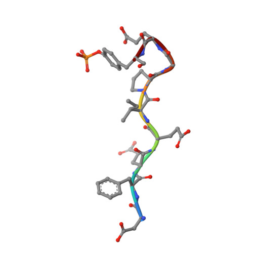
Compounds binding to the S2-S3 pockets of thrombin.

Nilsson, M.Hamalainen, M.Ivarsson, M.Gottfries, J.Xue, Y.Hansson, S.Isaksson, R.Fex, T.

(2009) J Med Chem 52: 2708-2715

  • DOI: https://doi.org/10.1021/jm8011849
  • Primary Citation of Related Structures:  
    3DA9

  • PubMed Abstract: 

    A set of compounds designed to bind to the S2-S3 pockets of thrombin was prepared. These compounds included examples with no interactions in the S1 pocket. Proline, a common P2 in many thrombin inhibitors, was combined with known P3 residues and P1 substituents of varying size and lipophilicity. Binding constants were determined using surface plasmon resonance (SPR) biosensor technology and were found to be in good agreement with results from an enzyme assay. A dramatic increase in affinity (100-1000 times) was seen for compounds incorporating an amino group capable of forming a hydrogen bond with gly216 in the protein backbone. The ligand efficiency was increased by including substituents that form stronger hydrophobic interactions with the P1 pocket. The binding mode was confirmed by X-ray analysis, which revealed the anticipated binding motif that included hydrogen bonds as well as a tightly bound water molecule. A QSAR model indicated that hydrogen bonding and lipophilicity were important for the prediction of binding constants. The results described here may have implications for how directed compound libraries for shallow protein pockets, like S2 and S3 in serine proteases, can be designed.


  • Organizational Affiliation

    School of Pure and Applied Natural Sciences, University of Kalmar, S-391 82 Kalmar, Sweden. Mikael.Nilsson@hik.se


Macromolecules
Find similar proteins by:  (by identity cutoff)  |  3D Structure
Entity ID: 1
MoleculeChains Sequence LengthOrganismDetailsImage
Thrombin light chain36Homo sapiensMutation(s): 0 
EC: 3.4.21.5
UniProt & NIH Common Fund Data Resources
Find proteins for P00734 (Homo sapiens)
Explore P00734 
Go to UniProtKB:  P00734
PHAROS:  P00734
GTEx:  ENSG00000180210 
Entity Groups  
Sequence Clusters30% Identity50% Identity70% Identity90% Identity95% Identity100% Identity
UniProt GroupP00734
Sequence Annotations
Expand
  • Reference Sequence
Find similar proteins by:  (by identity cutoff)  |  3D Structure
Entity ID: 2
MoleculeChains Sequence LengthOrganismDetailsImage
Thrombin heavy chain259Homo sapiensMutation(s): 0 
EC: 3.4.21.5
UniProt & NIH Common Fund Data Resources
Find proteins for P00734 (Homo sapiens)
Explore P00734 
Go to UniProtKB:  P00734
PHAROS:  P00734
GTEx:  ENSG00000180210 
Entity Groups  
Sequence Clusters30% Identity50% Identity70% Identity90% Identity95% Identity100% Identity
UniProt GroupP00734
Sequence Annotations
Expand
  • Reference Sequence

Find similar proteins by:  Sequence   |   3D Structure  

Entity ID: 3
MoleculeChains Sequence LengthOrganismDetailsImage
Hirudin peptideC [auth D]9N/AMutation(s): 0 
Sequence Annotations
Expand
  • Reference Sequence
Small Molecules
Ligands 2 Unique
IDChains Name / Formula / InChI Key2D Diagram3D Interactions
44U
Query on 44U

Download Ideal Coordinates CCD File 
E [auth B]beta-phenyl-D-phenylalanyl-N-propyl-L-prolinamide
C23 H29 N3 O2
HZKKJPDVZGOOPU-PZJWPPBQSA-N
NA
Query on NA

Download Ideal Coordinates CCD File 
D [auth B]SODIUM ION
Na
FKNQFGJONOIPTF-UHFFFAOYSA-N
Modified Residues  1 Unique
IDChains TypeFormula2D DiagramParent
PTR
Query on PTR
C [auth D]L-PEPTIDE LINKINGC9 H12 N O6 PTYR
Binding Affinity Annotations 
IDSourceBinding Affinity
44U BindingDB:  3DA9 Kd: 3100 (nM) from 1 assay(s)
IC50: 1.80e+4 (nM) from 1 assay(s)
PDBBind:  3DA9 Kd: 3100 (nM) from 1 assay(s)
Binding MOAD:  3DA9 Kd: 3100 (nM) from 1 assay(s)
Experimental Data & Validation

Experimental Data

  • Method: X-RAY DIFFRACTION
  • Resolution: 1.80 Å
  • R-Value Free: 0.215 
  • R-Value Work: 0.182 
  • R-Value Observed: 0.184 
  • Space Group: C 1 2 1
Unit Cell:
Length ( Å )Angle ( ˚ )
a = 69.83α = 90
b = 71.455β = 100.35
c = 71.905γ = 90
Software Package:
Software NamePurpose
REFMACrefinement
MAR345dtbdata collection
MOSFLMdata reduction
SCALAdata scaling
CCP4phasing

Structure Validation

View Full Validation Report


Ligand Structure Quality Assessment 


Entry History 

Deposition Data

Revision History  (Full details and data files)

  • Version 1.0: 2009-05-19
    Type: Initial release
  • Version 1.1: 2011-07-13
    Changes: Version format compliance
  • Version 1.2: 2024-04-03
    Changes: Data collection, Database references, Derived calculations, Refinement description