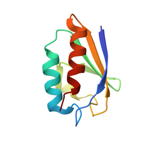
Structural investigation of a phosphorylation-catalyzed, isoaspartate-free, protein succinimide: crystallographic structure of post-succinimide His15Asp histidine-containing protein
Napper, S., Prasad, L., Delbaere, L.T.(2008) Biochemistry 47: 9486-9496
- PubMed: 18702519
- DOI: https://doi.org/10.1021/bi800847a
- Primary Citation of Related Structures:
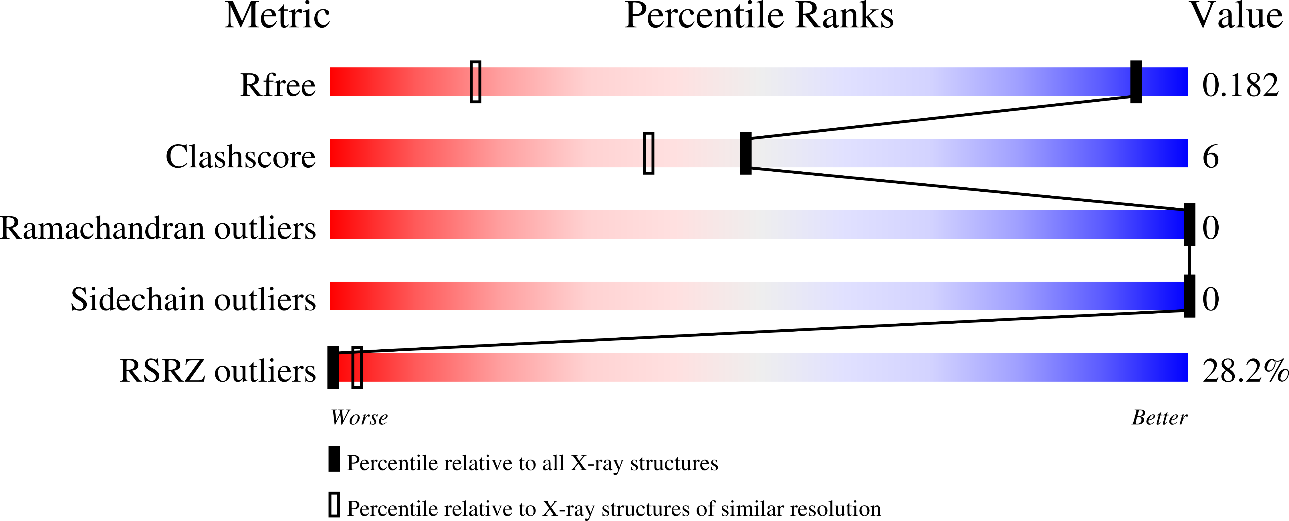
3CCD - PubMed Abstract:
Aspartates and asparagines can spontaneously cyclize with neighboring main-chain amides to form succinimides. These succinimides hydrolyze to a mixture of isoaspartate and aspartate products. Phosphorylation of aspartates is a common mechanism of protein regulation and increases the propensity for succinimide formation. Although typically regarded as a form of protein damage, we hypothesize succinimides could represent an effective mechanism of phosphoaspartate autophosphatase activity, provided hydrolysis is limited to aspartate products. We previously reported the serendipitous creation of a protein, His15Asp histidine-containing protein (HPr), which undergoes phosphorylation-catalyzed formation of a succinimide whose hydrolysis is seemingly exclusive for aspartate formation. Here, through the high-resolution structure of postsuccinimide His15Asp HPr, we confirm the absence of isoaspartate residues and propose mechanisms for phosphorylation-catalyzed succinimide formation and its directed hydrolysis to aspartate. His15Asp HPr represents the first characterized protein example of an isoaspartate-free succinimide and lends credence to the hypothesis that intramolecular cyclization could represent a physiological mechanism of autophosphatase activity. Furthermore, this indicates that current strategies for succinimide evaluation, based on isoaspartate detection, underestimate the frequencies of these reactions. This is considerably significant for evaluation of protein stability and integrity.
Organizational Affiliation:
Vaccine and Infectious Disease Organization, University of Saskatchewan, Saskatoon, Saskatchewan S7N 5E3, Canada. scott.napper@usask.ca