3C6G

Crystal structure of CYP2R1 in complex with vitamin D3


Experimental Data Snapshot

  • Method: X-RAY DIFFRACTION
  • Resolution: 2.80 Å
  • R-Value Free: 0.231 
  • R-Value Work: 0.193 
  • R-Value Observed: 0.195 

wwPDB Validation   3D Report Full Report


Ligand Structure Quality Assessment 


This is version 2.1 of the entry. See complete history


Literature

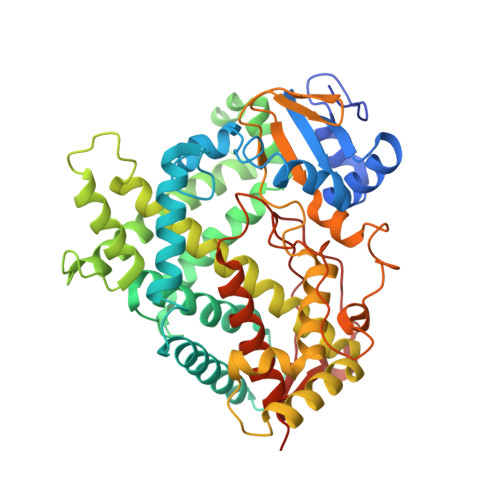
Structural analysis of CYP2R1 in complex with vitamin D3.

Strushkevich, N.Usanov, S.A.Plotnikov, A.N.Jones, G.Park, H.W.

(2008) J Mol Biol 380: 95-106

  • DOI: https://doi.org/10.1016/j.jmb.2008.03.065
  • Primary Citation of Related Structures:  
    3C6G

  • PubMed Abstract: 

    The activation of vitamin D to its hormonal form is mediated by cytochrome P450 enzymes. CYP2R1 catalyzes the initial step converting vitamin D into 25-hydroxyvitamin D. A CYP2R1 gene mutation causes an inherited form of rickets due to 25-hydroxylase deficiency. To understand the narrow substrate specificity of CYP2R1 we obtained the hemeprotein in a highly purified state, confirmed the enzyme as a vitamin D 25-hydroxylase, and solved the crystal structure of CYP2R1 in complex with vitamin D3. The CYP2R1 structure adopts a closed conformation with the substrate access channel being covered by the ordered B'-helix and slightly opened to the surface, which defines the substrate entrance point. The active site is lined by conserved, mostly hydrophobic residues. Vitamin D3 is bound in an elongated conformation with the aliphatic side-chain pointing toward the heme. The structure reveals the secosteroid binding mode in an extended active site and allows rationalization of the molecular basis of the inherited rickets associated with CYP2R1.


  • Organizational Affiliation

    Structural Genomics Consortium, University of Toronto, Toronto, Ontario, Canada.


Macromolecules
Find similar proteins by:  (by identity cutoff)  |  3D Structure
Entity ID: 1
MoleculeChains Sequence LengthOrganismDetailsImage
Cytochrome P450 2R1
A, B
479Homo sapiensMutation(s): 0 
Gene Names: CYP2R1
EC: 1.14.14
UniProt & NIH Common Fund Data Resources
Find proteins for Q6VVX0 (Homo sapiens)
Explore Q6VVX0 
Go to UniProtKB:  Q6VVX0
PHAROS:  Q6VVX0
GTEx:  ENSG00000186104 
Entity Groups  
Sequence Clusters30% Identity50% Identity70% Identity90% Identity95% Identity100% Identity
UniProt GroupQ6VVX0
Sequence Annotations
Expand
  • Reference Sequence
Oligosaccharides

Help

Entity ID: 2
MoleculeChains Length2D Diagram Glycosylation3D Interactions
Cycloheptakis-(1-4)-(alpha-D-glucopyranose)
C, D
7N/A
Glycosylation Resources
GlyTouCan:  G01435GL
GlyCosmos:  G01435GL
Small Molecules
Ligands 3 Unique
IDChains Name / Formula / InChI Key2D Diagram3D Interactions
HEM
Query on HEM

Download Ideal Coordinates CCD File 
H [auth A],
N [auth B]
PROTOPORPHYRIN IX CONTAINING FE
C34 H32 Fe N4 O4
KABFMIBPWCXCRK-RGGAHWMASA-L
VD3
Query on VD3

Download Ideal Coordinates CCD File 
I [auth A],
O [auth B]
(1S,3Z)-3-[(2E)-2-[(1R,3AR,7AS)-7A-METHYL-1-[(2R)-6-METHYLHEPTAN-2-YL]-2,3,3A,5,6,7-HEXAHYDRO-1H-INDEN-4-YLIDENE]ETHYLI DENE]-4-METHYLIDENE-CYCLOHEXAN-1-OL
C27 H44 O
QYSXJUFSXHHAJI-RWDMXNMGSA-N
UNX
Query on UNX

Download Ideal Coordinates CCD File 
E [auth A]
F [auth A]
G [auth A]
J [auth B]
K [auth B]
E [auth A],
F [auth A],
G [auth A],
J [auth B],
K [auth B],
L [auth B],
M [auth B]
UNKNOWN ATOM OR ION
X
Biologically Interesting Molecules (External Reference) 1 Unique
Experimental Data & Validation

Experimental Data

  • Method: X-RAY DIFFRACTION
  • Resolution: 2.80 Å
  • R-Value Free: 0.231 
  • R-Value Work: 0.193 
  • R-Value Observed: 0.195 
  • Space Group: C 2 2 21
Unit Cell:
Length ( Å )Angle ( ˚ )
a = 137.68α = 90
b = 163.051β = 90
c = 152.755γ = 90
Software Package:
Software NamePurpose
DENZOdata reduction
SCALEPACKdata scaling
MOLREPphasing
REFMACrefinement
PDB_EXTRACTdata extraction
ADSCdata collection

Structure Validation

View Full Validation Report


Ligand Structure Quality Assessment 


Entry History 

Revision History  (Full details and data files)

  • Version 1.0: 2008-02-19
    Type: Initial release
  • Version 1.1: 2011-07-13
    Changes: Version format compliance
  • Version 1.2: 2017-10-25
    Changes: Refinement description
  • Version 2.0: 2020-07-29
    Type: Remediation
    Reason: Carbohydrate remediation
    Changes: Atomic model, Data collection, Database references, Derived calculations, Non-polymer description, Structure summary
  • Version 2.1: 2023-08-30
    Changes: Data collection, Database references, Refinement description, Structure summary