


3C09
Crystal structure the Fab fragment of matuzumab (Fab72000) in complex with domain III of the extracellular region of EGFR
- PDB DOI: https://doi.org/10.2210/pdb3C09/pdb
- Classification: IMMUNE SYSTEM/TRANSFERASE
- Organism(s): Mus musculus, Homo sapiens
- Expression System: Mus musculus, Spodoptera frugiperda
- Mutation(s): No
- Deposited: 2008-01-18 Released: 2008-04-15
Experimental Data Snapshot
- Method: X-RAY DIFFRACTION
- Resolution: 3.20 Å
- R-Value Free: 0.299
- R-Value Work: 0.242
- R-Value Observed: 0.245

This is version 1.5 of the entry. See complete history.

Macromolecules
Find similar proteins by:
(by identity cutoff) | 3D Structure
Entity ID: 1 | |||||
|---|---|---|---|---|---|
| Molecule | Chains | Sequence Length | Organism | Details | Image |
| Matuzumab Fab Light chain | A [auth L], D [auth B] | 212 | Mus musculus | Mutation(s): 0 | 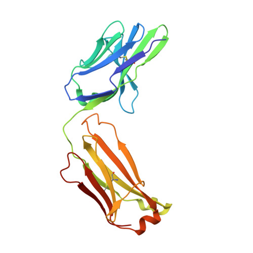 |
Entity Groups | |||||
| Sequence Clusters | 30% Identity50% Identity70% Identity90% Identity95% Identity100% Identity | ||||
Sequence AnnotationsExpand | |||||
| |||||
Find similar proteins by:
(by identity cutoff) | 3D Structure
Entity ID: 2 | |||||
|---|---|---|---|---|---|
| Molecule | Chains | Sequence Length | Organism | Details | Image |
| Matuzumab Fab Heavy chain | B [auth H], E [auth C] | 223 | Mus musculus | Mutation(s): 0 | 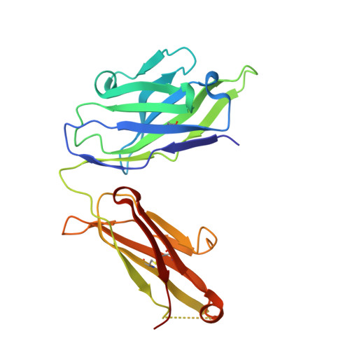 |
Entity Groups | |||||
| Sequence Clusters | 30% Identity50% Identity70% Identity90% Identity95% Identity100% Identity | ||||
Sequence AnnotationsExpand | |||||
| |||||
Find similar proteins by:
(by identity cutoff) | 3D Structure
Entity ID: 3 | |||||
|---|---|---|---|---|---|
| Molecule | Chains | Sequence Length | Organism | Details | Image |
| Epidermal growth factor receptor | C [auth A], F [auth D] | 214 | Homo sapiens | Mutation(s): 0 Gene Names: EGFR EC: 2.7.10.1 | 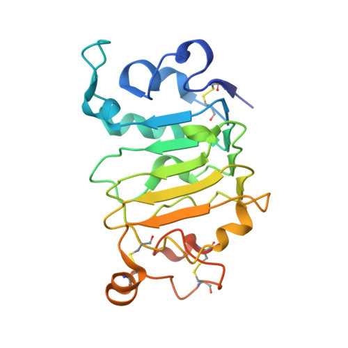 |
UniProt & NIH Common Fund Data Resources | |||||
Find proteins for P00533 (Homo sapiens) Explore P00533 Go to UniProtKB: P00533 | |||||
PHAROS: P00533 GTEx: ENSG00000146648 | |||||
Entity Groups | |||||
| Sequence Clusters | 30% Identity50% Identity70% Identity90% Identity95% Identity100% Identity | ||||
| UniProt Group | P00533 | ||||
Sequence AnnotationsExpand | |||||
| |||||
Small Molecules
| Ligands 3 Unique | |||||
|---|---|---|---|---|---|
| ID | Chains | Name / Formula / InChI Key | 2D Diagram | 3D Interactions | |
| NAG Query on NAG | G [auth A] H [auth A] K [auth A] L [auth A] M [auth A] | 2-acetamido-2-deoxy-beta-D-glucopyranose C8 H15 N O6 OVRNDRQMDRJTHS-FMDGEEDCSA-N |  | ||
| MAN Query on MAN | J [auth A] | alpha-D-mannopyranose C6 H12 O6 WQZGKKKJIJFFOK-PQMKYFCFSA-N |  | ||
| BMA Query on BMA | I [auth A], Q [auth D] | beta-D-mannopyranose C6 H12 O6 WQZGKKKJIJFFOK-RWOPYEJCSA-N |  | ||
Experimental Data & Validation
Experimental Data
- Method: X-RAY DIFFRACTION
- Resolution: 3.20 Å
- R-Value Free: 0.299
- R-Value Work: 0.242
- R-Value Observed: 0.245
- Space Group: C 1 2 1
Unit Cell:
| Length ( Å ) | Angle ( ˚ ) |
|---|---|
| a = 141.073 | α = 90 |
| b = 205.035 | β = 117.49 |
| c = 81.577 | γ = 90 |
| Software Name | Purpose |
|---|---|
| DENZO | data reduction |
| SCALEPACK | data scaling |
| PHASER | phasing |
| REFMAC | refinement |
| PDB_EXTRACT | data extraction |
Entry History
Deposition Data
- Released Date: 2008-04-15 Deposition Author(s): Ferguson, K.M., Schmiedel, J., Knoechel, T.
Revision History (Full details and data files)
- Version 1.0: 2008-04-15
Type: Initial release - Version 1.1: 2011-07-13
Changes: Non-polymer description, Version format compliance - Version 1.2: 2017-10-25
Changes: Refinement description - Version 1.3: 2019-12-25
Changes: Database references, Refinement description, Source and taxonomy - Version 1.4: 2020-07-29
Type: Remediation
Reason: Carbohydrate remediation
Changes: Data collection, Derived calculations, Structure summary - Version 1.5: 2023-11-01
Changes: Data collection, Database references, Refinement description, Structure summary
