3B6D

Crystal Structure of Streptomyces Cholesterol Oxidase H447Q/E361Q mutant (1.2A)


Experimental Data Snapshot

  • Method: X-RAY DIFFRACTION
  • Resolution: 1.20 Å
  • R-Value Free: 0.172 
  • R-Value Work: 0.132 
  • R-Value Observed: 0.132 

wwPDB Validation   3D Report Full Report


Ligand Structure Quality Assessment 


This is version 1.3 of the entry. See complete history


Literature

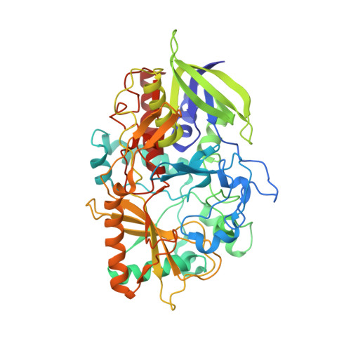
Distortion of flavin geometry is linked to ligand binding in cholesterol oxidase

Lyubimov, A.Y.Heard, K.Tang, H.Sampson, N.S.Vrielink, A.

(2007) Protein Sci 16: 2647-2656

  • DOI: https://doi.org/10.1110/ps.073168207
  • Primary Citation of Related Structures:  
    3B3R, 3B6D

  • PubMed Abstract: 

    Two high-resolution structures of a double mutant of bacterial cholesterol oxidase in the presence or absence of a ligand, glycerol, are presented, showing the trajectory of glycerol as it binds in a Michaelis complex-like position in the active site. A group of three aromatic residues forces the oxidized isoalloxazine moiety to bend along the N5-N10 axis as a response to the binding of glycerol in the active site. Movement of these aromatic residues is only observed in the glycerol-bound structure, indicating that some tuning of the FAD redox potential is caused by the formation of the Michaelis complex during regular catalysis. This structural study suggests a possible mechanism of substrate-assisted flavin activation, improves our understanding of the interplay between the enzyme, its flavin cofactor and its substrate, and is of use to the future design of effective cholesterol oxidase inhibitors.


  • Organizational Affiliation

    Department of Molecular Cell and Developmental Biology, University of California, Santa Cruz, California 95064, USA.


Macromolecules
Find similar proteins by:  (by identity cutoff)  |  3D Structure
Entity ID: 1
MoleculeChains Sequence LengthOrganismDetailsImage
Cholesterol oxidase504Streptomyces sp. SA-COOMutation(s): 2 
Gene Names: choA
EC: 1.1.3.6
UniProt
Find proteins for P12676 (Streptomyces sp. (strain SA-COO))
Explore P12676 
Go to UniProtKB:  P12676
Entity Groups  
Sequence Clusters30% Identity50% Identity70% Identity90% Identity95% Identity100% Identity
UniProt GroupP12676
Sequence Annotations
Expand
  • Reference Sequence
Small Molecules
Ligands 2 Unique
IDChains Name / Formula / InChI Key2D Diagram3D Interactions
FAE
Query on FAE

Download Ideal Coordinates CCD File 
C [auth A]FLAVIN-N7 PROTONATED-ADENINE DINUCLEOTIDE
C27 H34 N9 O15 P2
VWWQXMAJTJZDQX-UYBVJOGSSA-O
SO4
Query on SO4

Download Ideal Coordinates CCD File 
B [auth A]SULFATE ION
O4 S
QAOWNCQODCNURD-UHFFFAOYSA-L
Experimental Data & Validation

Experimental Data

  • Method: X-RAY DIFFRACTION
  • Resolution: 1.20 Å
  • R-Value Free: 0.172 
  • R-Value Work: 0.132 
  • R-Value Observed: 0.132 
  • Space Group: P 1 21 1
Unit Cell:
Length ( Å )Angle ( ˚ )
a = 51.3α = 90
b = 73.14β = 105.05
c = 63.14γ = 90
Software Package:
Software NamePurpose
REFMACrefinement
SHELXL-97refinement
CrystalCleardata collection
DENZOdata reduction
SCALEPACKdata scaling

Structure Validation

View Full Validation Report


Ligand Structure Quality Assessment 


Entry History 

Deposition Data

Revision History  (Full details and data files)

  • Version 1.0: 2007-12-18
    Type: Initial release
  • Version 1.1: 2011-07-13
    Changes: Source and taxonomy, Version format compliance
  • Version 1.2: 2021-11-10
    Changes: Database references, Derived calculations
  • Version 1.3: 2023-11-01
    Changes: Data collection, Refinement description