Structure-guided mutagenesis for the improvement of substrate specificity of Bacillus megaterium glucose 1-dehydrogenase IV
Nishioka, T., Yasutake, Y., Nishiya, Y., Tamura, T.(2012) FEBS J 279: 3264-3275
- PubMed: 22804868
- DOI: https://doi.org/10.1111/j.1742-4658.2012.08713.x
- Primary Citation of Related Structures:
3AUS, 3AUT, 3AY7 - PubMed Abstract:
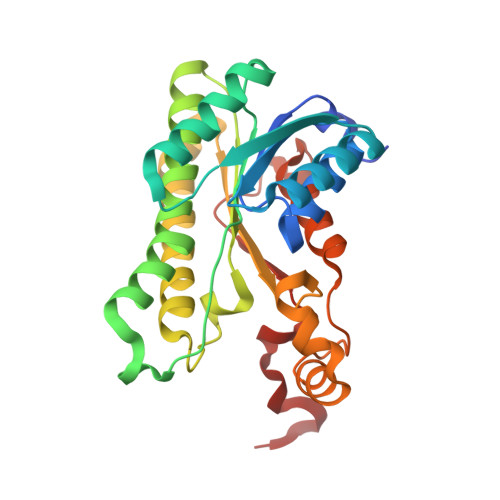
Bacillus megaterium IAM 1030 (Bacillus sp. JCM 20016) possesses four d-glucose 1-dehydrogenase isozymes (BmGlcDH-I, -II, -III and -IV) that belong to the short-chain dehydrogenase/reductase superfamily. The BmGlcDHs are currently used for a clinical assay to examine blood glucose levels. Of these four isozymes, BmGlcDH-IV has relatively high thermostability and catalytic activity, but the disadvantage of its broad substrate specificity remains to be overcome. Here, we describe the crystal structures of BmGlcDH-IV in ligand-free, NADH-bound and β-D-glucose-bound forms to a resolution of 2.0 Å. No major conformational differences were found among these structures. The structure of BmGlcDH-IV in complex with β-D-glucose revealed that the carboxyl group at the C-terminus, derived from a neighboring subunit, is inserted into the active-site pocket and directly interacts with β-D-glucose. A site-directed mutagenic study showed that destabilization of the BmGlcDH-IV C-terminal region by substitution with more bulky and hydrophobic amino acid residues greatly affects the activity of the enzyme, as well as its thermostability and substrate specificity. Of the six mutants created, the G259A variant exhibited the narrowest substrate specificity, whilst retaining comparable catalytic activity and thermostability to the wild-type enzyme.
Organizational Affiliation:
Graduate School of Agriculture, Hokkaido University, Sapporo, Japan.