3AR6

Calcium pump crystal structure with bound TNP-ADP and TG in the absence of calcium


Experimental Data Snapshot

  • Method: X-RAY DIFFRACTION
  • Resolution: 2.20 Å
  • R-Value Free: 0.287 
  • R-Value Work: 0.250 
  • R-Value Observed: 0.250 

wwPDB Validation   3D Report Full Report


Ligand Structure Quality Assessment 


This is version 1.3 of the entry. See complete history


Literature

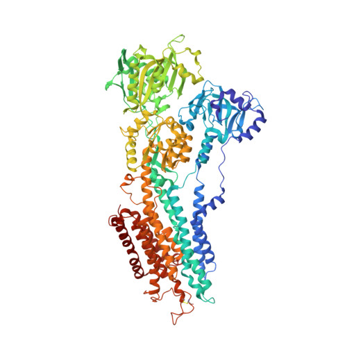
Trinitrophenyl derivatives bind differently from parent adenine nucleotides to Ca2+-ATPase in the absence of Ca2+.

Toyoshima, C.Yonekura, S.Tsueda, J.Iwasawa, S.

(2011) Proc Natl Acad Sci U S A 108: 1833-1838

  • DOI: https://doi.org/10.1073/pnas.1017659108
  • Primary Citation of Related Structures:  
    3AR2, 3AR3, 3AR4, 3AR5, 3AR6, 3AR7, 3AR8, 3AR9

  • PubMed Abstract: 

    Trinitrophenyl derivatives of adenine nucleotides are widely used for probing ATP-binding sites. Here we describe crystal structures of Ca(2+)-ATPase, a representative P-type ATPase, in the absence of Ca(2+) with bound ATP, trinitrophenyl-ATP, -ADP, and -AMP at better than 2.4-Å resolution, stabilized with thapsigargin, a potent inhibitor. These crystal structures show that the binding mode of the trinitrophenyl derivatives is distinctly different from the parent adenine nucleotides. The adenine binding pocket in the nucleotide binding domain of Ca(2+)-ATPase is now occupied by the trinitrophenyl group, and the side chains of two arginines sandwich the adenine ring, accounting for the much higher affinities of the trinitrophenyl derivatives. Trinitrophenyl nucleotides exhibit a pronounced fluorescence in the E2P ground state but not in the other E2 states. Crystal structures of the E2P and E2 ∼ P analogues of Ca(2+)-ATPase with bound trinitrophenyl-AMP show that different arrangements of the three cytoplasmic domains alter the orientation and water accessibility of the trinitrophenyl group, explaining the origin of "superfluorescence." Thus, the crystal structures demonstrate that ATP and its derivatives are highly adaptable to a wide range of site topologies stabilized by a variety of interactions.


  • Organizational Affiliation

    Institute of Molecular and Cellular Biosciences, University of Tokyo, Tokyo 113-0032, Japan. ct@iam.u-tokyo.ac.jp


Macromolecules
Find similar proteins by:  (by identity cutoff)  |  3D Structure
Entity ID: 1
MoleculeChains Sequence LengthOrganismDetailsImage
Sarcoplasmic/endoplasmic reticulum calcium ATPase 1995Oryctolagus cuniculusMutation(s): 1 
EC: 3.6.3.8
Membrane Entity: Yes 
UniProt
Find proteins for P04191 (Oryctolagus cuniculus)
Explore P04191 
Go to UniProtKB:  P04191
Entity Groups  
Sequence Clusters30% Identity50% Identity70% Identity90% Identity95% Identity100% Identity
UniProt GroupP04191
Sequence Annotations
Expand
  • Reference Sequence
Small Molecules
Ligands 5 Unique
IDChains Name / Formula / InChI Key2D Diagram3D Interactions
PTY
Query on PTY

Download Ideal Coordinates CCD File 
F [auth A],
G [auth A],
H [auth A]
PHOSPHATIDYLETHANOLAMINE
C40 H80 N O8 P
NJGIRBISCGPRPF-KXQOOQHDSA-N
TG1
Query on TG1

Download Ideal Coordinates CCD File 
D [auth A]OCTANOIC ACID [3S-[3ALPHA, 3ABETA, 4ALPHA, 6BETA, 6ABETA, 7BETA, 8ALPHA(Z), 9BALPHA]]-6-(ACETYLOXY)-2,3,-3A,4,5,6,6A,7,8,9B-DECAHYDRO-3,3A-DIHYDROXY-3,6,9-TRIMETHYL-8-[(2-METHYL-1-OXO-2-BUTENYL)OX Y]-2-OXO-4-(1-OXOBUTOXY)-AZULENO[4,5-B]FURAN-7-YL ESTER
C34 H50 O12
IXFPJGBNCFXKPI-FSIHEZPISA-N
12D
Query on 12D

Download Ideal Coordinates CCD File 
E [auth A]2',3'-O-[(1R,6R)-2,4,6-trinitrocyclohexa-2,4-diene-1,1-diyl]adenosine 5'-(trihydrogen diphosphate)
C16 H16 N8 O16 P2
WYOHYTDXVJMKHS-BENPRDFYSA-N
MG
Query on MG

Download Ideal Coordinates CCD File 
C [auth A]MAGNESIUM ION
Mg
JLVVSXFLKOJNIY-UHFFFAOYSA-N
NA
Query on NA

Download Ideal Coordinates CCD File 
B [auth A]SODIUM ION
Na
FKNQFGJONOIPTF-UHFFFAOYSA-N
Binding Affinity Annotations 
IDSourceBinding Affinity
TG1 BindingDB:  3AR6 Ki: 0.1 (nM) from 1 assay(s)
Kd: 0.2 (nM) from 1 assay(s)
IC50: min: 0.2, max: 3.9 (nM) from 2 assay(s)
Experimental Data & Validation

Experimental Data

  • Method: X-RAY DIFFRACTION
  • Resolution: 2.20 Å
  • R-Value Free: 0.287 
  • R-Value Work: 0.250 
  • R-Value Observed: 0.250 
  • Space Group: P 41 21 2
Unit Cell:
Length ( Å )Angle ( ˚ )
a = 71.619α = 90
b = 71.619β = 90
c = 588.192γ = 90
Software Package:
Software NamePurpose
BSSdata collection
CNSrefinement
DENZOdata reduction
SCALEPACKdata scaling
CNSphasing

Structure Validation

View Full Validation Report


Ligand Structure Quality Assessment 


Entry History 

Deposition Data

Revision History  (Full details and data files)

  • Version 1.0: 2011-02-02
    Type: Initial release
  • Version 1.1: 2011-07-13
    Changes: Version format compliance
  • Version 1.2: 2013-10-30
    Changes: Database references
  • Version 1.3: 2024-04-03
    Changes: Data collection, Database references, Derived calculations, Refinement description, Structure summary