High-resolution design of a protein loop.
Hu, X., Wang, H., Ke, H., Kuhlman, B.(2007) Proc Natl Acad Sci U S A 104: 17668-17673
- PubMed: 17971437
- DOI: https://doi.org/10.1073/pnas.0707977104
- Primary Citation of Related Structures:
2RB8, 2RBL - PubMed Abstract:
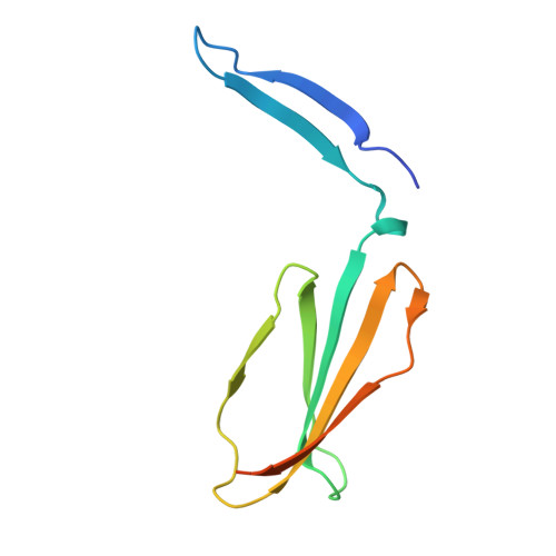
Despite having irregular structure, protein loops often adopt specific conformations that are critical to protein function. Most studies in de novo protein design have focused on creating proteins with regular elements of secondary structure connected by very short loops or turns. To design longer protein loops that adopt specific conformations, we have developed a protocol within the Rosetta molecular modeling program that iterates between optimizing the sequence and conformation of a loop in search of low-energy sequence-structure pairs. We have tested the procedure by designing 10-residue loops for the connection between the second and third strand in the beta-sandwich protein tenascin. Three low-energy designs from 7,200 flexible backbone trajectories were selected for experimental characterization. All three designs, called LoopA, LoopB, and LoopC, adopt stable folded structures. High-resolution crystal structures of LoopA and LoopB have been solved. LoopB adopts a structure very similar to the design model (0.46 A rmsd), and all but one of the side chains are modeled in the correct rotamers. LoopA crystallized at low pH in a structure that differs dramatically from our design model. It forms a strand-swapped dimer mediated by hydrogen bonds to protonated glutamic acids. Gel filtration indicates that the protein is not a dimer at neutral pH. These results suggest that the high-resolution design of protein loops is possible; however, they also highlight how small changes in protein energetics can dramatically perturb the low free energy structure of a protein.
Organizational Affiliation:
Department of Biochemistry and Biophysics, University of North Carolina, Chapel Hill, NC 27599, USA.