2ZJW

Crystal structure of human CK2 alpha complexed with Ellagic acid


Experimental Data Snapshot

  • Method: X-RAY DIFFRACTION
  • Resolution: 2.40 Å
  • R-Value Free: 0.274 
  • R-Value Work: 0.222 
  • R-Value Observed: 0.222 

wwPDB Validation   3D Report Full Report


Ligand Structure Quality Assessment 


This is version 1.2 of the entry. See complete history


Literature

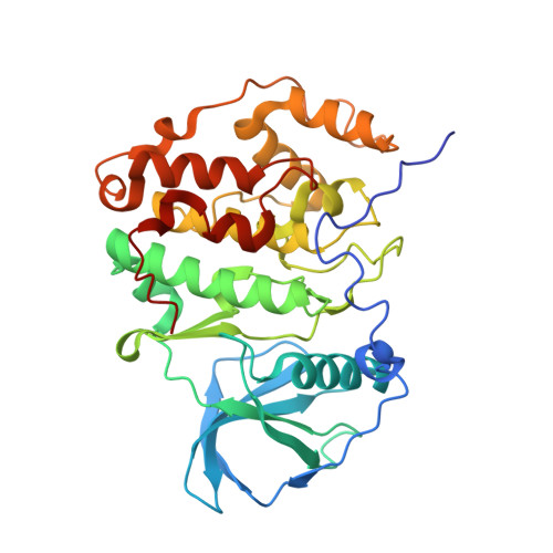
Structural insight into human CK2alpha in complex with the potent inhibitor ellagic acid

Sekiguchi, Y.Nakaniwa, T.Kinoshita, T.Nakanishi, I.Kitaura, K.Hirasawa, A.Tsujimoto, G.Tada, T.

(2009) Bioorg Med Chem Lett 19: 2920-2923

  • DOI: https://doi.org/10.1016/j.bmcl.2009.04.076
  • Primary Citation of Related Structures:  
    2ZJW

  • PubMed Abstract: 

    We determined the 2.35-A crystal structure of a human CK2 catalytic subunit (referred to as CK2alpha complexed with the ATP-competitive, potent CK2 inhibitor ellagic acid. The inhibitor binds to CK2alpha with a novel binding mode, including water-mediated hydrogen bonds. This structural information may support discovery of potent CK2 inhibitors.


  • Organizational Affiliation

    Graduate School of Science, Osaka Prefecture University, Sakai, Osaka, Japan.


Macromolecules
Find similar proteins by:  (by identity cutoff)  |  3D Structure
Entity ID: 1
MoleculeChains Sequence LengthOrganismDetailsImage
Casein kinase II subunit alpha340Homo sapiensMutation(s): 0 
EC: 2.7.11.1
UniProt & NIH Common Fund Data Resources
Find proteins for P68400 (Homo sapiens)
Explore P68400 
Go to UniProtKB:  P68400
PHAROS:  P68400
GTEx:  ENSG00000101266 
Entity Groups  
Sequence Clusters30% Identity50% Identity70% Identity90% Identity95% Identity100% Identity
UniProt GroupP68400
Sequence Annotations
Expand
  • Reference Sequence
Small Molecules
Ligands 1 Unique
IDChains Name / Formula / InChI Key2D Diagram3D Interactions
REF
Query on REF

Download Ideal Coordinates CCD File 
B [auth A]2,3,7,8-tetrahydroxychromeno[5,4,3-cde]chromene-5,10-dione
C14 H6 O8
AFSDNFLWKVMVRB-UHFFFAOYSA-N
Binding Affinity Annotations 
IDSourceBinding Affinity
REF PDBBind:  2ZJW Ki: 20 (nM) from 1 assay(s)
BindingDB:  2ZJW Ki: 20 (nM) from 1 assay(s)
IC50: 40 (nM) from 1 assay(s)
Binding MOAD:  2ZJW Ki: 20 (nM) from 1 assay(s)
Experimental Data & Validation

Experimental Data

  • Method: X-RAY DIFFRACTION
  • Resolution: 2.40 Å
  • R-Value Free: 0.274 
  • R-Value Work: 0.222 
  • R-Value Observed: 0.222 
  • Space Group: P 21 21 21
Unit Cell:
Length ( Å )Angle ( ˚ )
a = 47.953α = 90
b = 75.883β = 90
c = 78.421γ = 90
Software Package:
Software NamePurpose
HKL-2000data collection
MOLREPphasing
CNXrefinement
HKL-2000data reduction
HKL-2000data scaling

Structure Validation

View Full Validation Report


Ligand Structure Quality Assessment 


Entry History 

Deposition Data

Revision History  (Full details and data files)

  • Version 1.0: 2009-03-17
    Type: Initial release
  • Version 1.1: 2011-07-13
    Changes: Version format compliance
  • Version 1.2: 2023-11-01
    Changes: Data collection, Database references, Derived calculations, Refinement description