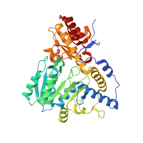
2Z9U

Crystal structure of pyridoxamine-pyruvate aminotransferase from Mesorhizobium loti at 2.0 A resolution


Experimental Data Snapshot

  • Method: X-RAY DIFFRACTION
  • Resolution: 2.00 Å
  • R-Value Free: 0.206 
  • R-Value Work: 0.156 
  • R-Value Observed: 0.158 

wwPDB Validation   3D Report Full Report


This is version 1.2 of the entry. See complete history


Literature

Crystal structure of pyridoxamine-pyruvate aminotransferase from Mesorhizobium loti MAFF303099

Yoshikane, Y.Yokochi, N.Yamasaki, M.Mizutani, K.Ohnishi, K.Mikami, B.Hayashi, H.Yagi, T.

(2008) J Biol Chem 283: 1120-1127

  • DOI: https://doi.org/10.1074/jbc.M708061200
  • Primary Citation of Related Structures:  
    2Z9U, 2Z9V, 2Z9W, 2Z9X

  • PubMed Abstract: 

    Pyridoxamine-pyruvate aminotransferase (PPAT; EC 2.6.1.30) is a pyridoxal 5'-phosphate-independent aminotransferase and catalyzes reversible transamination between pyridoxamine and pyruvate to form pyridoxal and L-alanine. The crystal structure of PPAT from Mesorhizobium loti has been solved in space group P4(3)2(1)2 and was refined to an R factor of 15.6% (R(free) = 20.6%) at 2.0 A resolution. In addition, the structures of PPAT in complexes with pyridoxamine, pyridoxal, and pyridoxyl-L-alanine have been refined to R factors of 15.6, 15.4, and 14.5% (R(free) = 18.6, 18.1, and 18.4%) at 1.7, 1.7, and 2.0 A resolution, respectively. PPAT is a homotetramer and each subunit is composed of a large N-terminal domain, consisting of seven beta-sheets and eight alpha-helices, and a smaller C-terminal domain, consisting of three beta-sheets and four alpha-helices. The substrate pyridoxal is bound through an aldimine linkage to Lys-197 in the active site. The alpha-carboxylate group of the substrate amino/keto acid is hydrogen-bonded to Arg-336 and Arg-345. The structures revealed that the bulky side chain of Glu-68 interfered with the binding of the phosphate moiety of pyridoxal 5'-phosphate and made PPAT specific to pyridoxal. The reaction mechanism of the enzyme is discussed based on the structures and kinetics results.


  • Organizational Affiliation

    Department of Bioresources Science, Faculty of Agriculture, Kochi University, Monobe-Otsu 200, Nankoku, Kochi 783-8502, Japan.


Macromolecules
Find similar proteins by:  (by identity cutoff)  |  3D Structure
Entity ID: 1
MoleculeChains Sequence LengthOrganismDetailsImage
Aspartate aminotransferase
A, B
392Mesorhizobium lotiMutation(s): 0 
Gene Names: mlr6806
EC: 2.6.1.30
UniProt
Find proteins for Q988B8 (Mesorhizobium japonicum (strain LMG 29417 / CECT 9101 / MAFF 303099))
Explore Q988B8 
Go to UniProtKB:  Q988B8
Entity Groups  
Sequence Clusters30% Identity50% Identity70% Identity90% Identity95% Identity100% Identity
UniProt GroupQ988B8
Sequence Annotations
Expand
  • Reference Sequence
Small Molecules
Ligands 2 Unique
IDChains Name / Formula / InChI Key2D Diagram3D Interactions
SO4
Query on SO4

Download Ideal Coordinates CCD File 
C [auth A]
D [auth A]
E [auth A]
H [auth B]
I [auth B]
C [auth A],
D [auth A],
E [auth A],
H [auth B],
I [auth B],
J [auth B]
SULFATE ION
O4 S
QAOWNCQODCNURD-UHFFFAOYSA-L
GOL
Query on GOL

Download Ideal Coordinates CCD File 
F [auth A],
G [auth A],
K [auth B],
L [auth B]
GLYCEROL
C3 H8 O3
PEDCQBHIVMGVHV-UHFFFAOYSA-N
Experimental Data & Validation

Experimental Data

  • Method: X-RAY DIFFRACTION
  • Resolution: 2.00 Å
  • R-Value Free: 0.206 
  • R-Value Work: 0.156 
  • R-Value Observed: 0.158 
  • Space Group: P 43 21 2
Unit Cell:
Length ( Å )Angle ( ˚ )
a = 68.545α = 90
b = 68.545β = 90
c = 311.635γ = 90
Software Package:
Software NamePurpose
REFMACrefinement
HKL-2000data collection

Structure Validation

View Full Validation Report


Entry History 

Deposition Data

Revision History  (Full details and data files)

  • Version 1.0: 2007-11-06
    Type: Initial release
  • Version 1.1: 2011-07-13
    Changes: Non-polymer description, Version format compliance
  • Version 1.2: 2023-11-01
    Changes: Data collection, Database references, Derived calculations, Refinement description